Correction: Human induced pluripotent stem cells derived neutrophils display strong anti-microbial potencies
Xing Hu, Baoqiang Kang, Mingquan Wang, Huaisong Lin, Zhiyong Liu, Zhishuai Zhang, Jiaming Gu, Yuchan Mai, Xinrui Guo, Wanli Ma, Han Yan, Shuoting Wang, Jingxi Huang, Junwei Wang, Jian Zhang, Tianyu Zhang, Bo Feng, Yanling Zhu, Guangjin Pan

Abstract
Genes, proteins, chemicals, diseases, species, mutations and cell lines named across the full text — each resolved to its canonical identifier and authoritative record.
Click any figure to enlarge with its caption.
 Figure 1
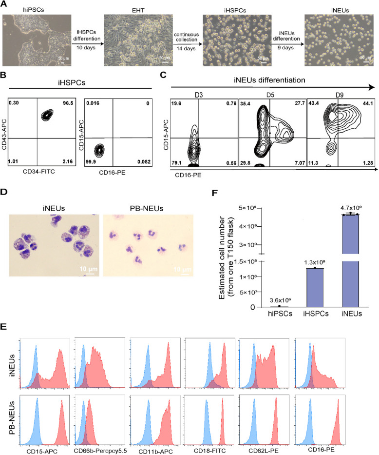 Figure 2
Peer Reviews
No public reviews on file for this paper yet. If you reviewed it on a platform where reviews are public (OpenReview, ICLR, NeurIPS, ICML), you can paste yours below so the community can read it here.
Videos
No videos yet. Explain this paper in a talk, walkthrough, or lecture? Add one.
Taxonomy
Topics3D Printing in Biomedical Research · Anesthesia and Neurotoxicity Research · Nanoplatforms for cancer theranostics
Correction: Cell Regeneration 14, 8 (2025)
https://doi.org/10.1186/s13619-025-00227-z
Following publication of the original article (Hu et al. 2025), the authors reported an error in Fig. 1E, the FACS data of surface markers CD11b and CD18 on iNEUs were mistakenly duplicated. Upon checking the original raw data, this error was caused by accidentally duplicating the same picture when formatting the figure.
The correct Fig. 1 has been provided in this Correction. These corrections do not affect the conclusions of this study.
The incorrect Fig. 1 is:
Fig. 1. Generation of neutrophils from hiPSCs. A Representative brightfield images of different stages of iNEUs differentiation from hiPSCs. Scale bar, 50 μm. B, C Representative FACS data of indicated markers on iHSPCs (B) and iNEUs differentiation at different time points (C). D Wright–Giemsa images of iNEUs and PB-NEUs. Scale bar, 10 μm. E Representative FACS data of indicated surface markers (CD15, CD66b, CD11b, CD18, CD62L and CD16; red filled) on iNEUs and PB-NEUs (blue, respective isotype control). F Estimated number of iHSPCs and iNEUs obtained from 3 × 10^6^ hiPSCs cultured in 1 T150 flask
The correct Fig. 1 is:
Fig. 1. Generation of neutrophils from hiPSCs. A Representative brightfield images of different stages of iNEUs differentiation from hiPSCs. Scale bar, 50 μm. B, C Representative FACS data of indicated markers on iHSPCs (B) and iNEUs differentiation at different time points (C). D Wright–Giemsa images of iNEUs and PB-NEUs. Scale bar, 10 μm. E Representative FACS data of indicated surface markers (CD15, CD66b, CD11b, CD18, CD62L and CD16; red filled) on iNEUs and PB-NEUs (blue, respective isotype control). F Estimated number of iHSPCs and iNEUs obtained from 3 × 10^6^ hiPSCs cultured in 1 T150 flask
The original article (Hu et al. 2025) has been corrected.