Transluminal forceps biopsy for a pancreaticojejunostomy stricture using a novel forceps biopsy device
Takeshi Ogura, Kimi Bessho, Nobuhiro Hattori, Yuki Uba, Hiroki Nishikawa

Abstract
Genes, proteins, chemicals, diseases, species, mutations and cell lines named across the full text — each resolved to its canonical identifier and authoritative record.
Click any figure to enlarge with its caption.
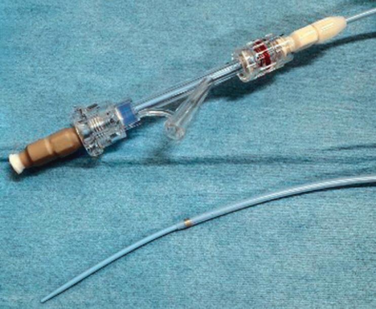 Fig. 1
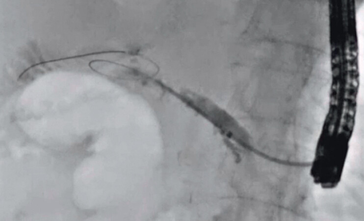 Fig. 2
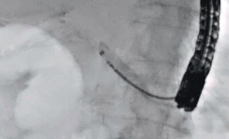 Fig. 3
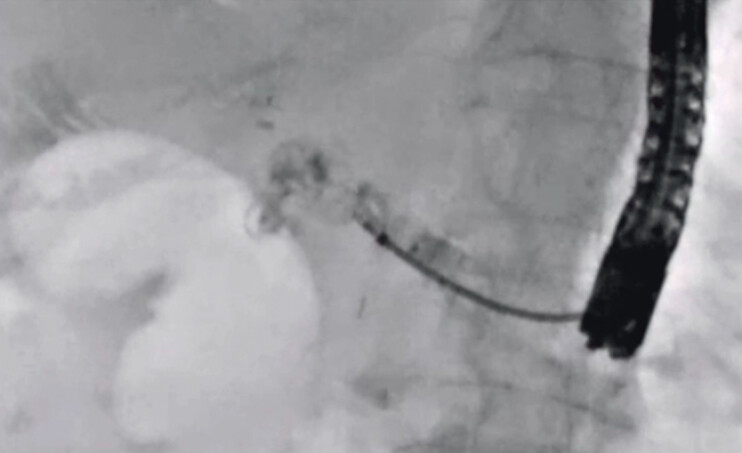 Fig. 4
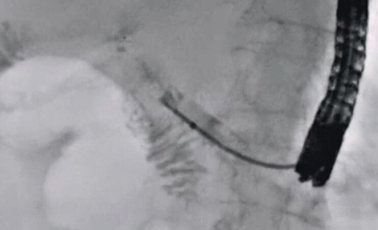 Fig. 5
Peer Reviews
No public reviews on file for this paper yet. If you reviewed it on a platform where reviews are public (OpenReview, ICLR, NeurIPS, ICML), you can paste yours below so the community can read it here.
Videos
No videos yet. Explain this paper in a talk, walkthrough, or lecture? Add one.
Taxonomy
TopicsPancreatic and Hepatic Oncology Research · Gallbladder and Bile Duct Disorders · Pancreatitis Pathology and Treatment
Endoscopic ultrasound (EUS)-guided pancreatic duct drainage (EUS-PDD) is indicated in cases of pancreatic duct obstruction, such as a pancreaticojejunostomy stricture, where endoscopic retrograde cholangiopancreatography (ERCP) has failed 1 2 3 4 5 . During the EUS-PDD procedure, leakage of pancreatic juice can occur until the stent has been deployed. Various procedures, such as stone removal or forceps biopsy, may increase the risk of adverse events. Transluminal biopsy may also be challenging, because matching the axes of the stricture site and the biopsy device is sometimes difficult, and there is a risk of puncturing the drainage tract. To overcome this, a novel forceps biopsy device has become available in Japan (ERCP Guide Sheath; Umidas, Kanagawa, Japan) ( Fig. 1 ). The diameter of the outer sheath of this device is 8.5 Fr, and the inner sheath is 6.0 Fr. In addition, the tip of this device is extremely tapered, conforming to a 0.035-inch guidewire. These characteristics allow penetration of stricture sites, and after removal of the inner sheath, various devices can be inserted. Side holes are also provided for the injection of contrast medium. This device can prevent pancreatic juice leakage and allow transluminal biopsy to be performed. Here, we describe transluminal biopsy in a pancreaticojejunostomy stricture.
The novel forceps biopsy device (ERCP Guide Sheath, Umidas, Kanagawa, Japan). The diameter of the outer sheath of this device is 8.5 Fr, and that of the inner sheath is 6.0 Fr. In addition, the tip of this device is extremely tapered, conforming to a 0.035-inch guidewire.
A 49-year-old man was admitted to our hospital due to pancreatitis caused by a pancreaticojejunostomy stricture. He had undergone pancreaticoduodenectomy to treat intraductal papillary mucinous carcinoma (IPMC) 10 months earlier. EUS-PDD was performed. After 2 weeks, to differentiate between recurrence of IPMC and benign stricture, transluminal biopsy was attempted. First, a 0.025-inch guidewire was deployed beside the EUS-PDD stent using an ERCP catheter. Then, the EUS-PDD stent was removed using a forceps biopsy device, and an ERCP guide sheath was inserted into the main pancreatic duct ( Fig. 2 ). The inner sheath was then removed, and it was possible to insert the forceps biopsy device smoothly into the main pancreatic duct through the ERCP guide sheath ( Fig. 3 ). To identify the stricture site, contrast medium was injected through the outer sheath ( Fig. 4 ). Transluminal forceps biopsy was then successfully performed ( Fig. 5 ), followed, finally, by stent deployment. The stricture was diagnosed histologically as a benign stricture ( Video 1 ).
The ERCP guide sheath is inserted into the main pancreatic duct.
The forceps biopsy device can be inserted smoothly into the main pancreatic duct through the ERCP guide sheath.
To identify the stricture site, contrast medium is injected through the outer sheath.
Transluminal forceps biopsy is successfully performed.
Transluminal forceps biopsy for a pancreaticojejunostomy stricture using a novel forceps biopsy device.Video 1
In conclusion, an ERCP guide sheath may be useful for forceps biopsy not only under ERCP, but also when using a transluminal approach.
Endoscopy_UCTN_Code_TTT_1AR_2AD
The reference list from the paper itself. Each links out to its DOI / PubMed record.
- 1Devière J Endoscopic ultrasound-guided pancreatic duct interventions Gastrointest Endosc Clin N Am 20233384585410.1016/j.giec.2023.04.00537709415 · doi ↗ · pubmed ↗
- 2Teh JL Teoh AYB Techniques and outcomes of endoscopic ultrasound guided-pancreatic duct drainage (EUS-PDD)J Clin Med 202312162610.3390/jcm 1204162636836161 PMC 9961828 · doi ↗ · pubmed ↗
- 3Giovannini MEUS-guided transenteric pancreatic duct drainage Best Pract Res Clin Gastroenterol 202210.1016/j.bpg.2022.10181536577534 · doi ↗ · pubmed ↗
- 4Ogura T Ohama H Higuchi K Endoscopic ultrasound-guided pancreatic transmural stenting and transmural intervention Clin Endosc 20205342943510.5946/ce.2019.13031771320 PMC 7403024 · doi ↗ · pubmed ↗
- 5Imoto A Ogura T Higuchi K Endoscopic ultrasound-guided pancreatic duct drainage: techniques and literature review of transmural stenting Clin Endosc 20205352553410.5946/ce.2020.17332967409 PMC 7548157 · doi ↗ · pubmed ↗