Pneumocephalus After Mediastinal Surgery
Mohamed Bhairis, Massine El Hammoumi, El Hassane Kabiri

Abstract
Genes, proteins, chemicals, diseases, species, mutations and cell lines named across the full text — each resolved to its canonical identifier and authoritative record.
Click any figure to enlarge with its caption.
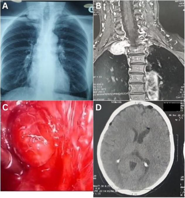 Figure 1
Peer Reviews
No public reviews on file for this paper yet. If you reviewed it on a platform where reviews are public (OpenReview, ICLR, NeurIPS, ICML), you can paste yours below so the community can read it here.
Videos
No videos yet. Explain this paper in a talk, walkthrough, or lecture? Add one.
Taxonomy
TopicsHead and Neck Surgical Oncology · Neurofibromatosis and Schwannoma Cases · Soft tissue tumor case studies
A 63-year-old female patient underwent investigation at a teaching hospital in Rabat, Morroco, in November 2023 for an incidental right apical opacity detected on a chest radiograph [Fig. 1A]. Magnetic resonance imaging (MRI) revealed a right apico-posterior mediastinal mass, which was excised via uniportal video-assisted thoracoscopic surgery (VATS) [Fig. 1B & Fig. 1C]. Histopathological analysis confirmed a diagnosis of schwannoma. Seventy-two hours after surgery, she developed a mild headache but exhibited no other neurological signs. An urgent computed tomography scan of the brain confirmed the presence of pneumocephalus [Fig. 1D]. Her symptoms resolved within 48 hours with bed rest and oxygen therapy. She was discharged without symptoms on the 10^th^ postoperative day.
1. Comments
Mediastinal schwannomas are benign neurogenic neoplasms with an excellent prognosis following surgical excision.^1^ VATS is a safe and effective approach for the resection of thoracic neurogenic tumours, except for dumbbell-shaped tumours.^2^ In our clinical practice, MRI serves as a cornerstone diagnostic modality for assessing the spatial relationship between the tumour and adjacent neurovascular structures, facilitating optimal surgical planning.^3^
Pneumocephalus may rarely occur as a postoperative complication. While often self-limiting and asymptomatic, conservative management is typically the preferred course of action. This approach should be complemented by vigilant clinical monitoring and appropriate follow-up to ensure complete resolution.^34^
Authors' Contribution
Mohamed Bhairis: Conceptualization, Writing - Original Draft. Massine El Hammoumi: Formal Analysis. El Hassane Kabiri: Writing - Original Draft, Writing - Review & Editing.
Ethics Statement
Written informed consent was obtained from the patient for publication of this case.
The reference list from the paper itself. Each links out to its DOI / PubMed record.
- 1Cingi E Emohare O Prielipp R. Major pneumocephalus after lung resection. A A Case Rep 2015; 4:68–70. https://doi.org/10.1213/XAA.0000000000000118.10.1213/XAA.000000000000011825774751 · doi ↗ · pubmed ↗
- 2Rakovich G Deslauriers J. Video-assisted and minimally invasive open chest surgery for the treatment of mediastinal tumours and masses. J Vis Surg 2017; 3:25. https://doi.org/10.21037/jovs.2017.01.01.10.21037/jovs.2017.01.0129078588 PMC 5638199 · doi ↗ · pubmed ↗
- 3Lee GS Lee MK Kim WJ Kim HS Kim JH Kim YS. Pneumocephalus and Pneumorrhachis due to a Subarachnoid Pleural Fistula That Developed after Thoracic Spine Surgery. Korean J Spine 2016; 13:164–6. https://doi.org/10.14245/kjs.2016.13.3.164.10.14245/kjs.2016.13.3.16427799999 PMC 5086471 · doi ↗ · pubmed ↗
- 4Asner S Chapuis-Taillard C Ris HB Gonzalez M. Pneumocephalus and pneumococcal meningitis after thoracic surgery. Asian Cardiovasc Thorac Ann 2011; 19:346–8. https://doi.org/10.1177/0218492311407796.10.1177/021849231140779622100930 · doi ↗ · pubmed ↗