What Happens If Hepatic Veins Drain to the Pulmonary Venous Atrium? Depends on the Physiology!
Kali A. Hopkins, Barry A. Love

TL;DR
Connecting hepatic veins to the pulmonary venous atrium can create blood shunts in the liver, with effects depending on the patient's physiology.
Contribution
Demonstrates how hepatic venous shunting varies based on physiology using two distinct clinical cases.
Findings
Hepatic venous connections between liver lobes can form significant shunts.
The direction of shunting depends on the patient's physiological state.
Two cases showed different hemodynamic outcomes from the same surgical connection.
Abstract
In rare instances in congenital cardiac surgery, ≥1 of the hepatic veins may end up incorporated either intentionally or inadvertently with the pulmonary venous atrium (oxygenated blood) while the remainder of the hepatic veins are left to drain normally with the systemic venous blood (less oxygenated blood). What happens when this is done? As it turns out, hepatic venous connections end up forming between the lobes of the liver, and a significant shunt results. The direction of shunting, however, depends on the physiology. We present 2 cases in which a single hepatic vein was left connected to the pulmonary venous atrium and produced very different hemodynamic consequences.
Genes, proteins, chemicals, diseases, species, mutations and cell lines named across the full text — each resolved to its canonical identifier and authoritative record.
Click any figure to enlarge with its caption.
 Figure 1
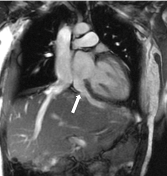 Figure 2
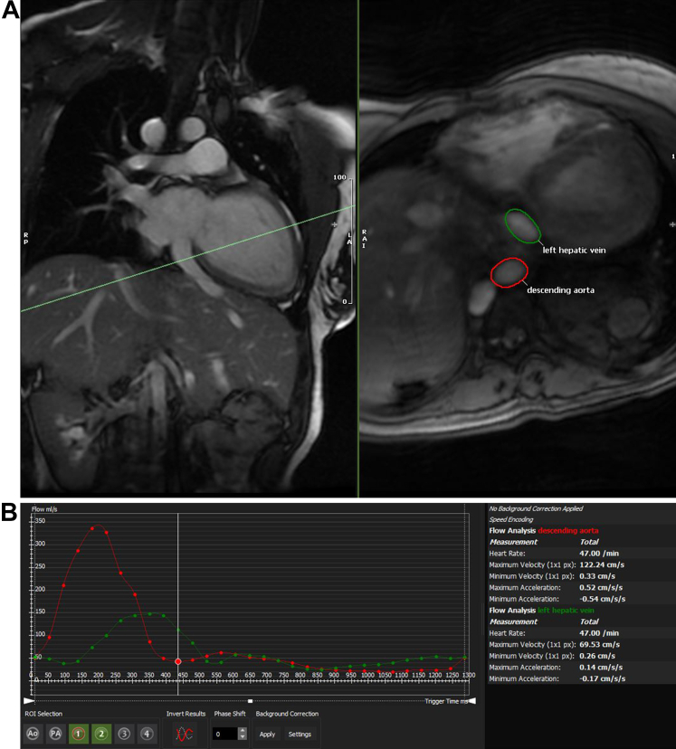 Figure 3
Peer Reviews
No public reviews on file for this paper yet. If you reviewed it on a platform where reviews are public (OpenReview, ICLR, NeurIPS, ICML), you can paste yours below so the community can read it here.
Videos
No videos yet. Explain this paper in a talk, walkthrough, or lecture? Add one.
Taxonomy
TopicsCongenital Heart Disease Studies · Pulmonary Hypertension Research and Treatments · Vascular anomalies and interventions