Reproducibility and reliability of flow quantification using CMR 2D-phase contrast and 4D-Flow in secondary mitral valve regurgitation
Yasaman Safarkhanlo, Martina Boscolo Berto, Giancarlo Spano, Benedikt Bernhard, Jonathan Schütze, Anselm W. Stark, Fabien Praz, Isaac Shiri, Alan A. Peters, Christof Schaub, Eva S. Peper, Chrysoula Garefa, Andreas Wahl, Jessica A. M. Bastiaansen, Christoph Gräni

TL;DR
This study compares the reliability of different MRI methods for measuring mitral valve regurgitation in older patients.
Contribution
The study identifies the 2D-PC standard method as the most reproducible for quantifying secondary mitral valve regurgitation using CMR.
Findings
The 2D-PC standard method showed the highest inter-reader and intra-reader reproducibility.
4D-flow methods had excellent intra-reader reliability but variable inter-reader agreement.
Standardized protocols and training could improve the clinical use of these MRI techniques.
Abstract
Accurate quantification of mitral valve regurgitation (MVR) is crucial for patient management. While different MVR quantification methods based on cardiac magnetic resonance imaging (CMR) exist, their reproducibility and reliability remain uncertain. This study aims to evaluate the reproducibility of different CMR 2D-phase contrast (PC) and 4D-flow MVR quantification methods. The inter-reader and intra-reader reproducibility were assessed using intraclass correlation coefficients (ICC). Seven methods were evaluated: 2D-PC standard (LVSV minus aortic flow), 2D-PC mitral-aortic (mitral inflow minus aortic flow), 2D-PC direct (quantifying mitral backflow), 4D-flow standard, 4D-flow mitral-aortic, 4D-flow direct, and volumetric method (LVSV minus RVSV) in 32 patients (74.8 ± 9.8 years, 28% females) with secondary MVR, analyzed independently by two experienced readers. A total of 26 patients…
Genes, proteins, chemicals, diseases, species, mutations and cell lines named across the full text — each resolved to its canonical identifier and authoritative record.
Click any figure to enlarge with its caption.
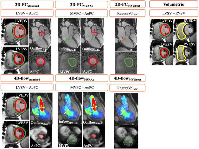 Figure 1
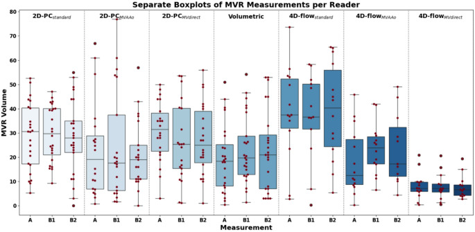 Figure 2
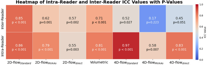 Figure 3
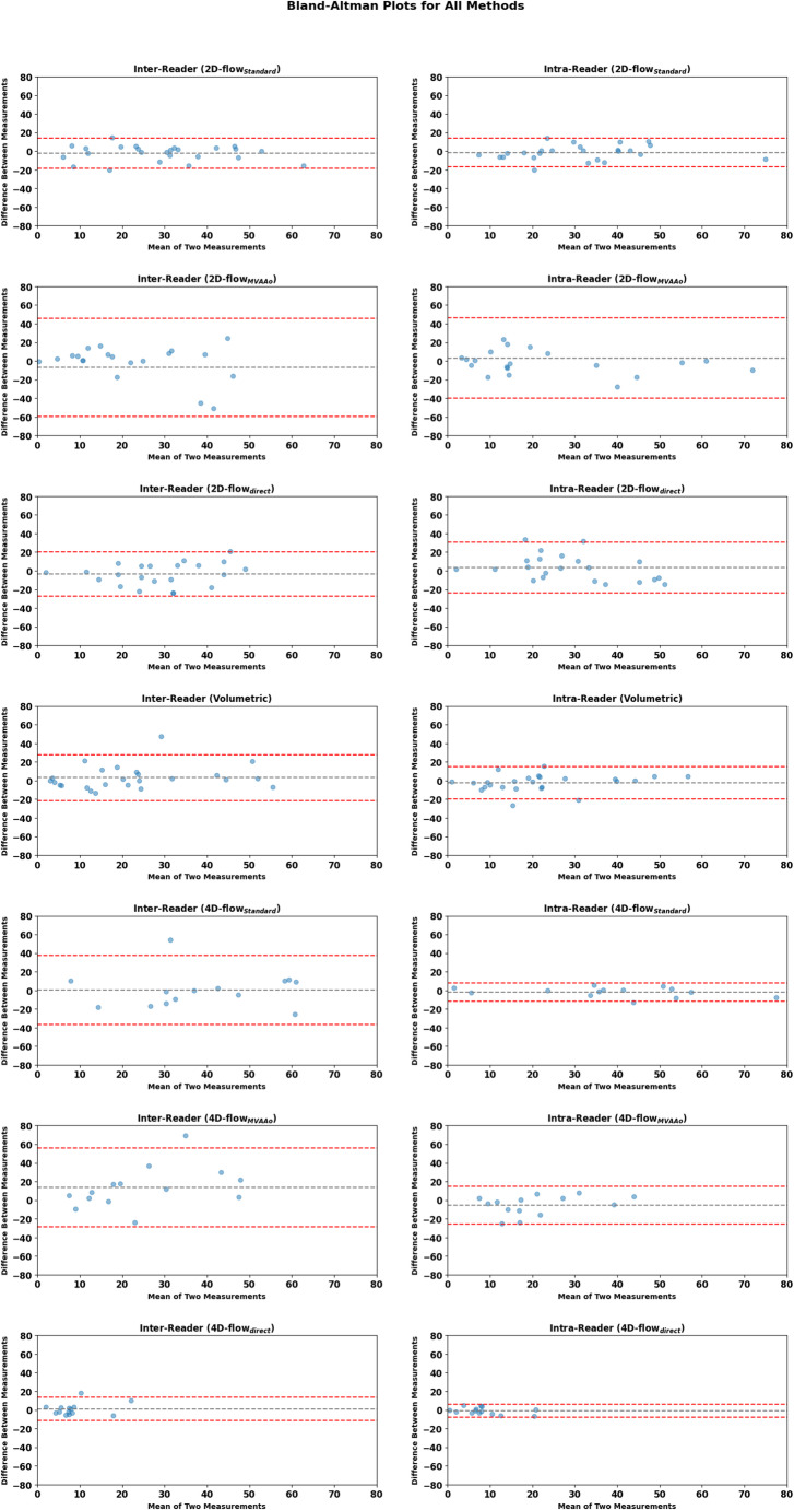 Figure 4
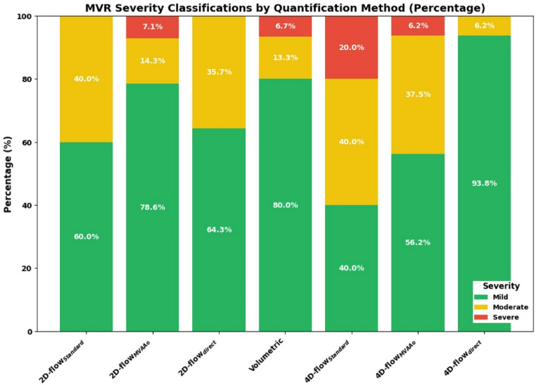 Figure 5
Peer Reviews
No public reviews on file for this paper yet. If you reviewed it on a platform where reviews are public (OpenReview, ICLR, NeurIPS, ICML), you can paste yours below so the community can read it here.
Videos
No videos yet. Explain this paper in a talk, walkthrough, or lecture? Add one.
Taxonomy
TopicsCardiac Valve Diseases and Treatments · Cardiac Imaging and Diagnostics · Advanced MRI Techniques and Applications