23Na MRI quantification of sodium content in porcine eyes after immersion in saltwater and freshwater en route to time in water estimation
Tobias Lindner, Adrian Konstantin Luyken, Chris Lappe, Oliver Stachs, Thoralf Niendorf, Matthias Lütgens, Stefan Polei, Brigitte Vollmar, Andreas Buettner, Sönke Langner, Marc-André Weber, Ebba Beller

TL;DR
This study explores using 23Na MRI to measure sodium in porcine eyes after immersion in saltwater or freshwater, aiming to estimate time in water and differentiate immersion types.
Contribution
The novel use of 23Na MRI for noninvasive sodium quantification in postmortem eyes to aid in forensic time-in-water estimation.
Findings
23Na MRI detected significant differences in vitreous body sodium concentration after 6 hours of immersion in saltwater versus freshwater.
Exponential curves modeled sodium concentration changes over immersion time in different saltwater and freshwater conditions.
Lens sodium concentrations also showed significant differences after 24 and 48 hours of immersion in various saltwater and freshwater solutions.
Abstract
Differentiation between saltwater and freshwater immersion as well as estimating the corpse’s time in water can be challenging. We aimed to establish and examine the feasibility of a novel approach based on sodium magnetic resonance imaging (23Na MRI) of the eye to facilitate noninvasive sodium quantification. Enucleated porcine eyes were immersed in NaCl 0.9%, NaCl 3.0%, NaCl 5.85%, distilled water (DW) or lake water (LW) at different time intervals, followed by 23Na 7-T MRI sodium quantification. After 6 h of immersion, a significant difference in vitreous body (VB) sodium concentration was found for NaCl 5.85% versus DW or LW (p ≤ 0.019). After 24 and 48 h of immersion, a significant difference in VB sodium concentration was found for NaCl 5.85% versus DW, LW, NaCl 3.0% or NaCl 0.9%, as well as for NaCl 3.0% versus DW, LW or NaCl 0.9% (p ≤ 0.001). After 24 h of immersion, lens…
Genes, proteins, chemicals, diseases, species, mutations and cell lines named across the full text — each resolved to its canonical identifier and authoritative record.
Click any figure to enlarge with its caption.
 Figure 1
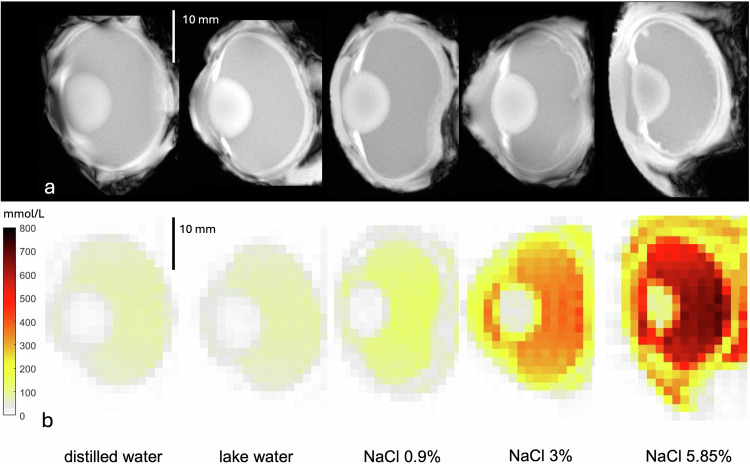 Figure 2
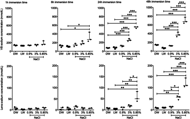 Figure 3
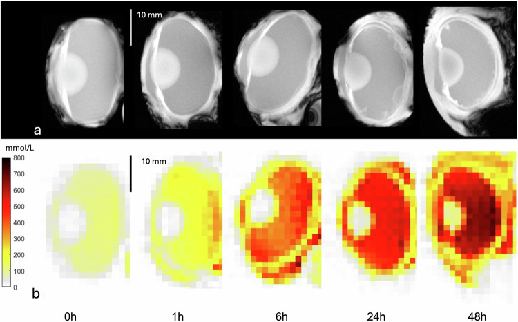 Figure 4
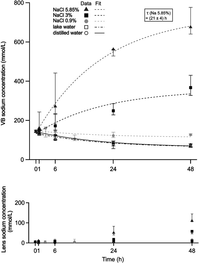 Figure 5
 Figure 6
Peer Reviews
No public reviews on file for this paper yet. If you reviewed it on a platform where reviews are public (OpenReview, ICLR, NeurIPS, ICML), you can paste yours below so the community can read it here.
Videos
No videos yet. Explain this paper in a talk, walkthrough, or lecture? Add one.
Taxonomy
TopicsAutopsy Techniques and Outcomes · Forensic Anthropology and Bioarchaeology Studies · Radiation Dose and Imaging