A new genus and two new species of Empoascini (Hemiptera, Cicadellidae, Typhlocybinae) from China

Abstract
Genes, proteins, chemicals, diseases, species, mutations and cell lines named across the full text — each resolved to its canonical identifier and authoritative record.
Click any figure to enlarge with its caption.
 Figures 1–6
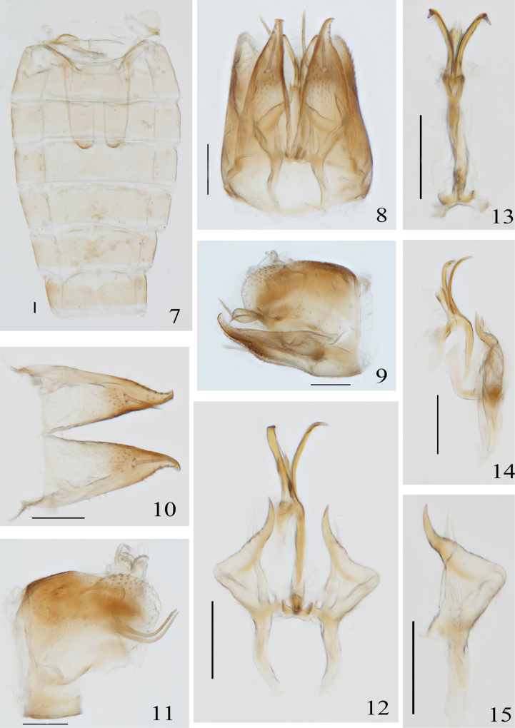 Figures 7–15
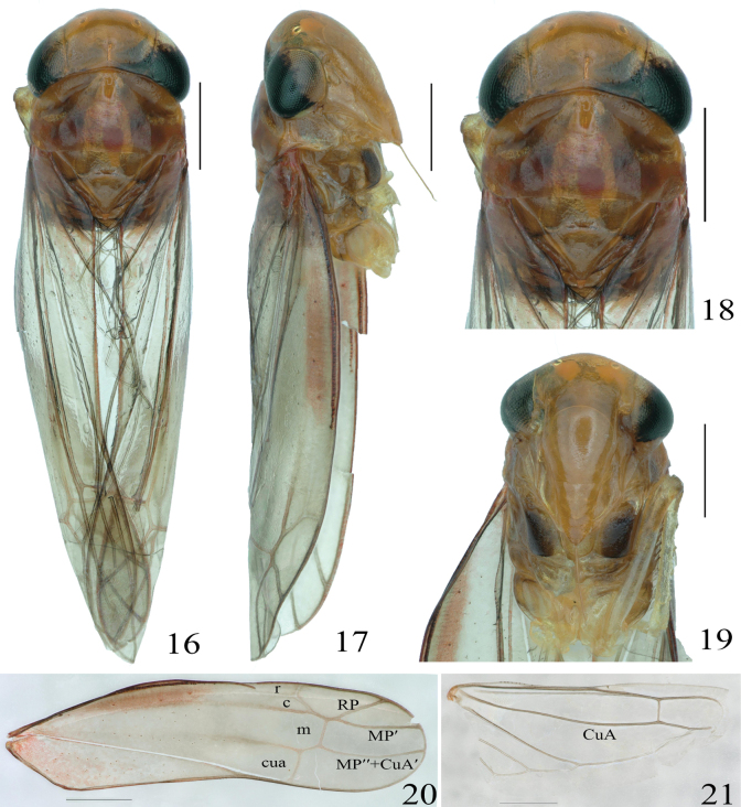 Figures 16–21
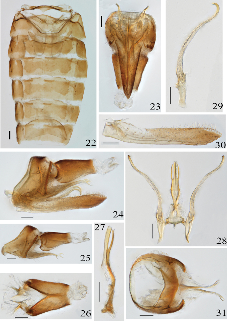 Figures 22–31
| 1 | Apex of ventral pygofer appendage unbranched |
|
| – | Apex of ventral pygofer appendage branched |
|
| 2 | Aedeagus with two processes |
|
| – | Aedeagus shaft base only with a forked process |
|
| 3 | The basal processes of aedeagus shaft located on both sides of the shaft, symmetrical, not exceeding shaft length |
|
| – | The basal processes of aedeagus shaft are asymmetrical and unequal in length |
|
Peer Reviews
No public reviews on file for this paper yet. If you reviewed it on a platform where reviews are public (OpenReview, ICLR, NeurIPS, ICML), you can paste yours below so the community can read it here.
Videos
No videos yet. Explain this paper in a talk, walkthrough, or lecture? Add one.
Taxonomy
TopicsPhytoplasmas and Hemiptera pathogens · Genomics and Phylogenetic Studies · Cocoa and Sweet Potato Agronomy
Introduction
Species of the tribe Empoascini (Hemiptera, Cicadellidae, Typhlocybinae) are major pests of agriculture and forestry (Chen, 1979; Qin et al. 2014). Currently, approximately 107 genera and 1396 species of Empoascini have been reported worldwide, while in China, 50 genera and 262 species are known (Xu et al. 2019, 2021a, 2023; Yu et al. 2020; Wang et al. 2021; Dmitriev et al. 2022; Ding et al. 2024).
The genus Velu was initially established by Ghauri in 1964, with the type species V.caricae Ghauri being sourced from India. Subsequently, Dworakowska (1980) and Sohi and Mann (1986) each added a new species within this genus, and Zhang and Qin (2004a) compiled a comprehensive species list and formulated a taxonomic key for three newly discovered species of Velu from China. To date, six species of Velu have been reported across the Eastern Oceanic and Palaearctic realms.
In the current study, a new genus with a new species, are described. In addition, one species of Velu is also described from Yunnan, thereby augmenting the existing knowledge of the genus within the Chinese region and contributing to the broader understanding of its global diversity.
Material and methods
The morphological terminology adopted in this work was in accordance with the standards set by Southern (1982), Zhang (1990), Dworakowska (1993), Dietrich (2005), and Xu et al. (2021b). External features of specimens were meticulously recorded and the body length, measured from the apex of the vertex to the tip of the wing, was precisely determined using a KEYENCE VHX-6000 digital camera. Additionally, the male genitalia were photographed with high clarity employing a Nikon Eclipse Ni-E compound microscope. All type specimens of the newly described species have been permanently deposited at the Institute of Entomology, Guizhou University, located in Guiyang, China (GUGC).
Taxonomy
Singulus
Taxon classificationAnimaliaHemipteraCicadellidae
Yao & Yu gen. nov.
BE80194B-C335-5844-8246-22995CDCB973
https://zoobank.org/DE522CBC-D4B2-46A3-A1A3-D5C347071500
Type species.
Singulusfurcatus Yao & Yu, sp. nov. here designated.
Diagnosis.
The new genus is similar to Alafrasca Lu & Qin, 2014, Lumicella Lu & Qin, 2013, Schizandrasca Anufriev, 1972, and Circinans Qin & Liu, 2014 in that the CuA veins of hindwings are branched (Fig. 6), but the new genus differs from the above genera by the following characters: the subgenital plate is triangular with a broad base and only one large seta (Figs 8, 10). Alafrasca Lu & Qin differs from the new genus in lacking an anal process branching at the apex (Lu and Qin 2014b). Lumicella Lu & Qin differs from the new genus in the following aspects: the subgenital plate is not triangular, the paramere is slim, and the apophysis bears a prominent dentifer (Lu et al. 2013). Schizandrasca Anufriev, 1972 differs from the new genus in lacking a paired aedeagus processes (Anufriev, 1972a). Circinans Qin & Liu differs by the anal process with a broad and extended caudad (Lu and Qin 2014a).
Description.
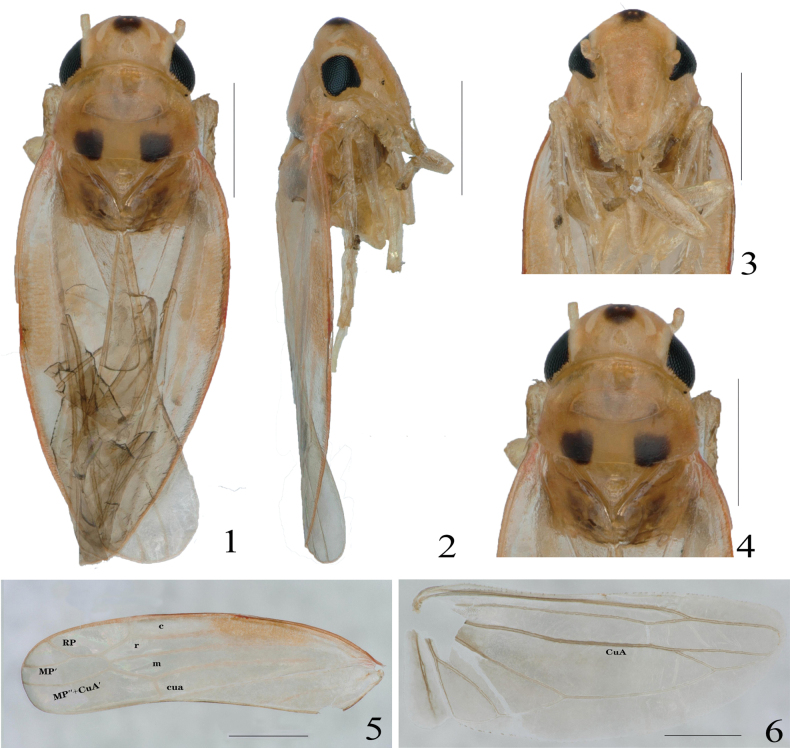
Body slender (Figs 1–4). Crown nearly as wide as pronotum, middle length of crown less than distance between eyes, a black spot in middle of vertex, with ocelli. Coronal suture present and not reaching anterior margin of crown. Pronotum with anterior margin arcuate, angles on both sides of mesoscutellum are black, scutoscutellar sulcus not reaching both margins (Figs 1, 4). Face larger than wide, elevated in lateral view (Figs 2, 3). Forewing RP and MP′ veins stalked at base, arising from r cell, r cell longest, r cell and m cell subequal in width, 1^st^ apical cell largest, followed by 4^th^ apical, 3^rd^ apical triangular, which is about one-third of wing length (Fig. 5). Hindwing CuA vein branched, and branch point located on or at intersection of CuA and MP′′ veins (Fig. 6).
Abdominal apodemes bursts, slender, parallel (Fig. 7). Male genitalia broad, pygofer without process, posterior margin bluntly rounded, with fine setae (Figs 8, 9). Subgenital plate triangular, broad at base, narrowing to tip, with 1 macrosetae (Fig. 10). Apex of paramere is apical, fine setae distributed in subapical, tip without teeth (Figs 12, 15). Aedeagus curved in lateral view, with paired process, without dorsal apodeme, preatrium well developed (Figs 12, 13). Anal process developed, slender, curved toward apex in an arc, exceeding posterior margin of pygofer, proximal with an indentation (Figs 9, 11). Connective not developed (Figs 12, 13).
Singulusfurcatus Yao & Yu, sp. nov., male holotype. 1. Habitus, dorsal view; 2. Habitus, lateral view; 3. Face, anterior view; 4. Head and pronotum, dorsal view; 5. Forewing; 6. Hindwing. Scale bars: 500 μm.
Etymology.
The new genus is named after the subgenital plate of males with only 1 macroseta.
Distribution.
Oriental.
Singulus
furcatus
Taxon classificationAnimaliaHemipteraCicadellidae
Yao & Yu sp. nov.
B1E66CDB-0061-5776-A015-603F309C9703
https://zoobank.org/96411AF4-525D-4621-9E8A-79762C9AE9A1
Description.
Body red (Figs 1–4). Anterior margin of crown arcuate, posterior margin concave, a large black spot in middle of anterior margin of crown. Eyes black, ocellus located on top of head in line with antennae, and crown suture conspicuous, not reaching anterior margin of crown. Pronotum red, anterior area with irregular spots, and angles on both sides of scutellum have black spots (Figs 1, 4). Forewings reddish and hindwings translucent (Figs 5, 6).
Singulusfurcatus Yao & Yu, sp. nov. 7. Abdominal apodemes; 8. Male genitalia, ventral view; 9. Male genitalia, left lateral view; 10. Subgenital plate; 11. Pygofer, pygofer appendage and anal tube, lateral view; 12. Aedeagus, paramere and connective, dorsal view; 13. Aedeagus and connective, dorsal view; 14. Aedeagus and connective, left lateral view; 15. Paramere. Scale bars: 100 μm.
Male abdominal apodemes extend to middle of 5^th^ abdominal segment, and they are slender and parallel (Fig. 7). Lateral valves of male pygofer side broad at base, narrowed in terminal half, coarsely setae, without ventral appendage (Fig. 9). Anal process developed, extending toward tip, distinctly exceeding posterior margin of pygofer, subterminally with many grooves (Fig. 9). Subgenital plate gradually narrowed basally to tip, with 1 macrosetae and 17 marginal setae (Figs 9, 10). Paramere curved, inflated at middle, pointed at tip and without teeth (Figs 12, 15). Aedeagus curved in lateral view, with short whiskers at apex of shaft and with a pair of protuberances on each side of ventral margin of shaft, which are distinctly curved ventrally beyond apex of shaft, and in dorsal view with shaft protuberances forked (Figs 12, 13, 14). Connective not developed (Figs 12, 13).
Material examined.
Holotype: • ♂, Mengla, Yunnan, 2015-May-14, coll. Bin Yan. Paratypes: • 2♂, same as holotype; 1♂, Dading Mountain, Guangdong, August 11–13, 2006, coll. Zhong-Hui Zhou.
Etymology.
The name of the new species derives from the shaft having a pair of protuberances, which are distinctly forked at the tips when viewed dorsally.
Measurement.
Length of male 2.7–2.9 mm.
Distribution.
China (Yunnan, Guangdong).
Velu
Taxon classificationAnimaliaHemipteraCicadellidae
Genus
Ghauri, 1964
141544F5-B248-52B5-B39B-01E4776F113A
Velu Ghauri, 1964e: 467.
Type species.
Velucaricae Ghauri, 1964.
Diagnosis.
Body robust (Figs 16–19). The anterior margin of the vertex is arc-shaped, and the posterior margin is concave, nearly parallel to each other. The width of the vertex is almost equal to the thorax, and its median length is less than the width between the eyes. Coronal suture is prominent, extending to the middle of vertex, with shallow depressions on both sides; the lateral margin of the pronotum has an inverted trapezoid transverse concavity (Figs 16, 18). Forewing RP and MP’ veins healed at base, and the terminal veins all originate from the m cell, which is wider and longer than the c cell and the r cell. The 3^rd^ apical is triangular, and the submarginal veins of the 2^nd^ apical are nearly parallel (Fig. 20). Hindwing CuA veins are unbranched (Fig. 21).
Male abdominal apodemes are well developed and parallel (Fig. 22). Pygofer appendage elongate (Fig. 31). Subgenital plate arranged a row of macrosetae (Fig. 30). Aedeagus has a preatrium, with a process at the base of shaft in ventral view or not (Fig. 27). Paramere slender, curved in a semicircle at the apex, and fine setae are present on the inner margin at the curved portion (Figs 28, 29).
Distribution.
Oriental and Palaearctic.
Checklist and distribution of Velu Ghauri, 1964
1. Veluclaustrum Yao & Yang, sp. nov.
Distribution. China (Yunnan).
2. Velucaricae Ghauri, 1964
Velucaricae Ghauri, 1964e: 468.
Distribution. Pakistan, India (Karnataka, West Bengal).
3. Velupruthii Dworakowska, 1980
Velupruthii Dworakowska, 1980: 155.
Distribution. India (Karnataka).
4. Veluantelopus Sohi & Mann, 1986
Veluantelopus Sohi & Mann, 1986: 100.
Distribution. India.
5. Velufurcatum Zhang & Qin, 2004
Velufurcatum Zhang & Qin, 2004: 276.
Distribution. China (Yunnan).
6. Velulongiprojectum Zhang & Qin, 2004
Velulongiprojectum Zhang & Qin, 2004: 277.
Distribution. China (Yunnan).
7. Velupleuroprominens Zhang & Qin, 2004
Velupleuroprominens Zhang & Qin, 2004: 278.
Distribution. China (Yunnan).
Key to species (males) of Velu Ghauri from China
**: **
Velu
claustrum
Taxon classificationAnimaliaHemipteraCicadellidae
Yao & Yang sp. nov.
18BC040A-5D62-52FB-A233-C14081AE02E8
https://zoobank.org/876ACE3F-9A03-493A-A415-B7B006AE54AF
Description.
Whole body orange-yellow (Figs 16–19). Anterior margin of head curved, posterior margin concave, ocelli brown at junction of head and face, eyes light brown, and crown suture conspicuous, reaching to middle of crown. Pronotum with anterior margin arcuate, anterior domain with a transverse dark depression, middle and posterior domains orange, elevated, and intershield grooves brown, not reaching both margins (Figs 16, 18). Face broad, frontclyeal area elevated in lateral view (Figs 17, 19). Forewing RP and MP’ veins heal at base, m-cells wider than and longer than c cells and r cells, 3^rd^ apical triangular, 2^nd^ apical lateral veins subparallel (Fig. 20). Hindwing CuA veins unbranched (Fig. 21).
Veluclaustrum Yao & Yang, sp. nov., male holotype. 16. Habitus, dorsal view; 17. Habitus, lateral view; 18. Head and pronotum, dorsal view; 19. Face, anterior view; 20. Forewing; 21. Hindwing. Scale bars: 500 μm.
Veluclaustrum Yao & Yang, sp. nov. 22. Abdominal apodemes; 23. Male genitalia, ventral view; 24. Male genitalia, left lateral view; 25. Pygofer, pygofer appendage and anal tube, lateral view; 26. Anal tube; 27. Aedeagus, left lateral view; 28. Aedeagus, paramere and connective, ventral view; 29. Paramere; 30. Subgenital plate; 31. Pygofer and pygofer appendage. Scale bars: 100 μm.
Male abdominal apodemes extending to 4^th^ abdominal segment (Fig. 22). Pygofer side lateral view basally broad, apex membranous, dorsal margin abruptly narrowed near middle, resulting in large difference in width between terminal and basal pygofer side lateral view, and pygofer appendage long, distinctly exceeding apex of pygofer, apex split (Figs 23, 24, 25, 31). Anal process absent (Fig. 26). Subgenital plate long, base slightly broad, subequal in width in terminal half, with 8 individual macrosetae, 39 long, fine setae (Fig. 30). Paramere long and narrow, hooked and curved in terminal half, with fine setae subterminally (Figs 28, 29). Aedeagus curved in lateral view, with a pair of lateral processes at base of shaft, which are slightly shorter and narrower than shaft, while apex of paramere curved towards shaft in ventral view (Figs 27, 28). Connective base broad, terminal margin deeply concave (Figs 27, 28).
Material examined.
Holotype: • 1♂, Menghai, Xishuangbanna, Yunnan, 2013-July-14, coll. Ji-Chun Xing. Paratypes: • 1♂, 4♀, same collecting information as holotype.
Etymology.
The new species is named after the pair of dark-coloured striped depressions on both margins of the pronotum in specimens examined.
Measurement.
Length of males 3.9–4.0 mm, females 4.1–4.5 mm.
Remarks.
The new species is similar to V.pleuroprominens Zhang & Qin, 2004 in having a well-developed preatrium, slightly shorter than the shaft, and symmetrical lateral processes of the shaft (Fig. 28). The latter species is different from the former in that: (1) the apex of the two lateral processes at the base of the shaft deviates from the shaft; (2) the apex of the pygofer of the abdominal processes is unbranched (Zhang & Qin, 2004a).
Distribution.
China (Yunnan).
Supplementary Material
XML Treatment for Singulus
XML Treatment for Singulus furcatus
XML Treatment for Velu
XML Treatment for Velu claustrum
The reference list from the paper itself. Each links out to its DOI / PubMed record.
- 1Anufriev GA (1972 a) New and little known Palaearctic genera and species of Typhlocybinae (Homoptera: Cicadellidae). Bulletin de l’Académie Polonaise des Sciences.Série des Sciences Biologiques 20(1): 35–42.
- 2Chen ZM (1979) Composition and succession of disease and pest in tea garden. Chinese Tea (1): 6–8. [in Chinese]
- 3Dietrich CH (2005) Keys to the families of Cicadomorpha and subfamilies and tribes of Cicadellidae (Hemiptera: Auchenorrhyncha). The Florida Entomologist 88(4): 502–517. 10.1653/0015-4040(2005)88[502:KTTFOC]2.0.CO;2 · doi ↗
- 4Ding Y Yan B Yu XF Yang MF (2024) A new genus and three new species of Empoascini (Hemiptera: Cicadellidae: Typhlocybinae) from China.Zootaxa 5433(1): 121–132. 10.11646/zootaxa.5433.1.539645761 · doi ↗ · pubmed ↗
- 5Dmitriev DA Angelova R Anufriev GA Bartlett CR Blanco-Rodríguez E Borodin OI Cao YH Cara C Deitz LL Dietrich CH Dmitrieva MO El-Sonbati SA Evangelista de Souza O Gjonov IV Gonçalves AC Gonçalves CC Hendrix SV Mc Kamey S Kohler M Kunz G MalenovskýI Morris BO Novoselova M Pinedo-Escatel JA Rakitov RA Rothschild MJ Sanborn AF Takiya DM Wallace MS Zahniser JN (2022) [onward] Empoascini Distant, 1908. World Auchenorrhyncha Database. Taxon Pages. [Retrieved on 2025-03-26] https://hoppers.speciesfile.org/otus/946156/overview
- 6Dworakowska I (1980) On some Typhlocybinae from India (Homoptera: Auchenorrhyncha: Cicadellidae).Entomologische Abhandlungen und Berichte aus dem Staatlichen Museum fur Tierkunde in Dresden 43(8): 151–201. 10.1515/9783112653227-010 · doi ↗
- 7Dworakowska I (1993) Remarks on Alebra Fieb. and Eastern Hemisphere Alerini (Auchenorrhyncha: Cicadellidae: Typhlocybinae).Entomotaxonomia 15(2): 91–121.
- 8Ghauri MSK (1964) New fig leaf-hoppers (Homoptera: Cicadelloidea) from India with redescription of allied species under new genera. Annals and Magazine of Natural History (Series 13) 6: 465–475. [1963] 10.1080/00222936308651384 · doi ↗