Correlation between Acetowhite Examination, Dermoscopy, and Histopathology in Patients with Anogenital Warts
Qaira Anum, Vesri Yossy, Nellia Fonna

TL;DR
This study compares non-invasive methods like acetowhite examination and dermoscopy to histopathology for diagnosing anogenital warts, finding that dermoscopy is highly accurate.
Contribution
The study provides a direct comparison of diagnostic accuracy between acetowhite examination, dermoscopy, and histopathology for anogenital warts.
Findings
Acetowhite examination had 91% sensitivity but low specificity and NPV.
Dermoscopy showed 100% sensitivity and specificity, making it highly accurate.
Dermoscopy can reduce the need for invasive histopathological procedures.
Abstract
Anogenital warts, caused by human papillomavirus (HPV) infection, are characterized by papular lesions in the anogenital region. Acetowhite examination and dermoscopy are non-invasive methods that might aid in confirming the clinical diagnosis. This study aimed to evaluate the diagnostic accuracy of acetowhite examination and dermoscopy compared to histopathological findings in anogenital warts. This cross-sectional study included patients diagnosed with anogenital warts at Dr. M. Djamil Padang Hospital (Padang, Indonesia) from January 2023 to December 2023. Using the purposive sampling method, 62 lesions from 54 patients (28 men and 26 women) aged 16-59 years were analyzed. Each lesion underwent acetowhite examination, dermoscopy, and histopathological examination. Descriptive analyses were performed on subject characteristics and dermoscopic features, while sensitivity and specificity…
Genes, proteins, chemicals, diseases, species, mutations and cell lines named across the full text — each resolved to its canonical identifier and authoritative record.
Click any figure to enlarge with its caption.
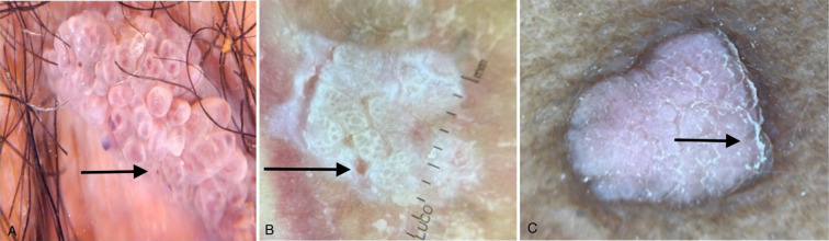 Figure 1
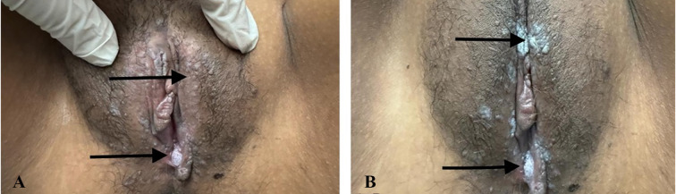 Figure 2
| Category | Subcategory | Frequency N (%) |
|---|---|---|
| Age | 16-25 | 21 (39) |
| 26-35 | 23 (42) | |
| 36-45 | 7 (13) | |
| 46-55 | 2 (4) | |
| 56-59 | 1 (2) | |
| Total | 54 (100) | |
| Sex | Male | 28 (52) |
| Female | 26 (48) | |
| Total | 54 (100) | |
| Lesion Location | Urethra | 1 (2) |
| Penis | 10 (18) | |
| Scrotum | 7 (13) | |
| Vulva | 21 (39) | |
| Pubis | 2 (4) | |
| Perineum | 10 (18) | |
| Perianal | 24 (44) | |
| Dermoscopy Image | Location | Total Lesions (n=62) | |
|---|---|---|---|
| Genitals (n=37) | Perianal (n=25) | ||
| Unspecified | 2 (5%) | 0 | 2 (3%) |
| Fingerlike | 23 (63%) | 18 (72%) | 41 (66%) |
| Knoblike | 7 (19%) | 3 (12%) | 10 (16%) |
| Mosaics | 2 (5%) | 2 (8%) | 4 (7%) |
| Fingerlike/Knoblike | 2 (5%) | 0 | 2 (3%) |
| Knoblike/Mosaic | 0 | 1 (4%) | 1 (2%) |
| Fingerlike/Mosaic | 1 (3%) | 1 (4%) | 2 (3%) |
| Total | 37 | 25 | 62 (100%) |
Peer Reviews
No public reviews on file for this paper yet. If you reviewed it on a platform where reviews are public (OpenReview, ICLR, NeurIPS, ICML), you can paste yours below so the community can read it here.
Videos
No videos yet. Explain this paper in a talk, walkthrough, or lecture? Add one.
Taxonomy
TopicsNeonatal skin health care
What’s Known
- Anogenital warts, caused by human papillomavirus (HPV) infection, present as papular lesions or warts in the anogenital area. Acetowhite examination and dermoscopy are non-invasive tools used to confirm the clinical diagnosis.
- Previous studies demonstrated that acetowhite examination had high sensitivity and specificity, while dermoscopy exhibited high specificity but lower sensitivity for diagnosing anogenital warts.
What’s New
- This study provides a comprehensive assessment of the diagnostic accuracy of acetowhite examination and dermoscopy compared to histopathological findings in anogenital warts.
- Analyzing 62 lesions from 54 patients, the study identified fingerlike and knoblike patterns as the most frequent dermoscopic features. While both acetowhite examination and dermoscopy showed high sensitivity (91.0% and 100%, respectively), dermoscopy achieved 100% specificity, highlighting its superior diagnostic reliability for positive cases.
Introduction
Condyloma acuminatum (CA) or anogenital warts, is a manifestation of human papillomavirus (HPV) infection in the anogenital region. It is estimated that 9-13% of the global population experiences CA, with the highest prevalence occurring between the ages of 20 and 39 years. ^ 1 , 2 ^ Based on data from the Dermatology and Venereology Polyclinic of Dr. M. Djamil General Hospital, anogenital warts ranked as the most frequently treated condition in the sexually transmitted infections division in 2017. ^ 3 ^
A definite diagnosis of anogenital warts typically requires biopsy and histopathological examination, as the gold standard, followed by HPV serotype testing. However, these methods are time-consuming and costly. Dermoscopy, a non-invasive diagnostic examination tool, has been increasingly utilized to support the diagnosis of non-tumor and non-pigmented skin disorders. A study by Yeh and others demonstrated that dermoscopic imaging had high sensitivity and accuracy, achieving a sensitivity of 97.4% and a specificity of 87.5% when compared to histopathology. ^ 4 ^
The diagnosis of condylomata acuminata is primarily based on clinical observations. For ambiguous lesions, the acetowhite test can be performed on suspected areas, with a 10 to 15-min waiting period. This test reveals white discoloration in HPV-affected regions, aiding in diagnosis and guiding protective measures using keratolytic agents, although it may lack specificity. Biopsy is generally reserved for atypical lesions, uncertain diagnoses, lack of improvement with standard treatment, or worsening conditions during therapy. ^ 5 ^ While previous studies highlighted the potential of non-invasive methods, such as acetowhite testing and dermoscopy to enhance diagnostic accuracy, further research is required to establish their reliability in clinical practice. This study aimed to evaluate the effectiveness of acetowhite and dermoscopy examinations in correlation with histopathological results for patients with anogenital warts.
Patients and Methods
This cross-sectional study employed a purposive sampling method. The patients who were diagnosed with anogenital warts at Dr. M. Djamil Hospital, Padang, Indonesia, were enrolled from January 2023 to December 2023. The inclusion criteria included patients with a confirmed diagnosis of anogenital warts by a dermatologist based on the clinical features and histopathological findings, as well as those willing to participate in the study. The exclusion criteria included patients with other medical conditions, such as additional sexually transmitted diseases, that could affect the results, as well as those who did not provide consent.
The sample size was calculated using Cochran’s formula to ensure that the research findings were reliable and had adequate statistical power. Patients undergoing treatments such as immunotherapy or other therapies that could influence diagnostic results, or those with medical conditions impacting the examination, were given special consideration.
The study procedure involved five key steps:
1- History Taking: Interviews were conducted to gather detailed information about the patient’s medical history
2- Physical Examination: A thorough examination was performed to identify and assess anogenital wart lesions.
3- Acetowhite examination: Acetic acid solution (Merck Indo Avidatama, Indonesia) was applied to evaluate the acetowhite reactions in the lesions.
4- Dermoscopic Examination: A dermatoscope (DermLite DL3N, DermLite LLC headquartered in Aliso Viejo, California, USA) was used to observe the clinical features of the lesions.
Histopathological Examination: A biopsy was performed using histopathology chemicals (Sigma-Aldrich, USA) and biopsy instruments (Bard Inc., USA) for definitive diagnosis, as histopathology is considered the gold standard for diagnosing anogenital warts. ^ 6 ^
Ethical Considerations
This study was approved by the Health Research Ethics Committee of Dr. M. Djamil Hospital, Padang, Andalas University, Indonesia, with the approval code L.B.02.2/57/435/2023. Written informed consent was obtained from all participants before the examination.
Statistical Analysis
Data analysis was conducted using two approaches.
Descriptive Analysis: subject characteristics, clinical findings, and dermoscopic features of anogenital warts were analyzed descriptively to provide an overview of patient demographics and lesion characteristics.
Diagnostic Accuracy: a 2×2 table was used to compare the results of acetowhite examination, dermoscopy examination, and histopathology examinations. Sensitivity and specificity were calculated using predetermined formulas, enabling researchers to evaluate the diagnostic accuracy of each method for anogenital warts. ^ 5 ^
A 2×2 table was used to compare the results of acetowhite examination, dermoscopy, and histopathology. Sensitivity, specificity, positive predictive value (PPV), negative predictive value (NPV), and likelihood ratios (LR^+^ and LR^-^) were calculated using the following formulas:
-Sensitivity: [A/(A+C)]×100%
-Specificity: [A/(A+B)]×100%
-NPV: [D/(D+B)]×100%
-PPV: [A/(A+C)]×100%
-
LR^+^: [A/(A+B)]×100%
-
LR^-^: [D/(D+C)]×100%
Results
A total of 62 lesions from 54 patients (28 men and 26 women) aged 16-59 years were studied (table 1). The CA lesions were found in multiple locations. In male patients, lesions were observed in the scrotum (n=7, 13%), perianal area (n=24, 44%), and penis (n=10, 18%). In female patients, lesions were located in the vulva (n=21, 39%), perianal area (n=24, 44%), and perineum (n=10, 18%) (table 1).
The distribution of dermoscopic features is summarized in table 2. The fingerlike pattern was the most common feature (66%), followed by the knoblike pattern (16%), mosaic pattern (7%), unspecified patterns (3%), and combined fingerlike/knoblike patterns (3%). Representative images of the fingerlike, knoblike, and mosaic patterns are shown in figures 1A, B, and C, respectively.
(A) Dermoscopic images of condyloma acuminata show a finger-like pattern, characterized by papillomatous structures with fingerlike projections and prominent, smooth, organized papular lesions. (B) Knoblike pattern, marked by larger round lesions, resembles buttons with uniform length and diameter. (C) The mosaic pattern shows characteristics of condyloma acuminata (Dermlite DL3N).
Based on lesion location, the sensitivity of dermoscopy for genital lesions was 3% for unspecified patterns, 27% for fingerlike patterns, 10% for knoblike patterns, 3% for mosaic patterns, 3% for fingerlike/knoblike patterns, and 2% for fingerlike/mosaic patterns. The specificity of dermoscopy for genital lesions was 100%. For perianal lesions, the sensitivity and specificity of dermoscopy examination were as follows: fingerlike patterns (22% sensitivity, 100% specificity), knoblike patterns (5% sensitivity, 100% specificity), mosaic patterns (3% sensitivity, and 100% specificity), knoblike/mosaic patterns (2% sensitivity, 100% specificity), and fingerlike/mosaic patterns (2% sensitivity, 100% specificity), LR+[A/(A+B)]×100% and LR-[D/(D+C)]×100%. The acetowhite examination results showed 49 positive and 5 negative cases, with a sensitivity of 100%, specificity of 100%, and PPV of 100% (figure 2).
The clinical presentation of a patient with condyloma acuminata typically includes the presence of papular or wart-like lesions in the anogenital region. (A) Warts before the application of 5% acetic acid (black arrow); (B) Warts after the application of 5% acetic acid, shows white staining (black arrow).
In this study, we evaluated the suitability of acetowhite examination and dermoscopy compared to histopathology results in patients with anogenital warts. Histopathological examination of 54 patients revealed flat layered epithelium characterized by hyperkeratosis, parakeratosis, acanthosis, hyperplasia, papillomatosis, and koilocytosis. Additionally, lymphocytes, plasma cells, and PMN leukocytes were observed in the connective tissue stroma.
The analysis demonstrated different levels of sensitivity and specificity for the diagnostic methods. The Acetowhite examination showed a sensitivity of 91.0% and a PPV of 91.0%. However, specificity and NPV could not be calculated, indicating limitations in identifying negative results. In contrast, dermoscopy exhibited 100% sensitivity and specificity, along with a high PPV. However, NPV could not be calculated due to the absence of negative results. Overall, both acetowhite examination and dermoscopy were effective in detecting condyloma acuminata. In contrast, the inability to calculate specificity and NPV for acetowhite examination highlighted the need for further verification of negative results.
Discussion
In this study, acetowhite examination demonstrated a sensitivity of 91% and a PPV of 91%, but specificity could not be calculated. Dermoscopy, on the other hand, exhibited 100% sensitivity, specificity, and PPV. These findings were in agreement with previous research by Zhu and others who reported a sensitivity of 92.30% and specificity of 58.20% for anal warts using dermoscopy. ^ 2 ^
In this study, perianal warts lesions were divided into six dermoscopic patterns: unspecific, fingerlike, knoblike, mosaic, knoblike/mosaic, and fingerlike/mosaic. The sensitivity and specificity of each type were compared with the histopathology results.
Dermoscopic examination of genital and perianal lesions showed high specificity for diagnosing anogenital condyloma acuiminata (ACA), suggesting that dermoscopy can serve as a valuable diagnostic tool. However, it could not replace histopathological examination as the gold standard.
The acetowhite test, which involves applying a 3-5% aqueous solution of acetic acid, is a relatively reliable diagnostic method. The acetowhitening effect is attributed to the coagulation of epithelial cytokeratins, particularly cytokeratin 10. However, this test has limitations, as false-positive results can occur, particularly in inflammatory conditions. Despite this, a strong correlation was reported between histopathological findings indicative of HPV and the presence of HPV DNA in acetowhite areas. ^ 9 ^
Variations in acetowhiteness among different wart types might stem from differences in epidermal hydration. In hyperplastic anogenital warts, the moist epithelium facilitates acetic acid penetration, leading to protein (cytokeratin) coagulation and the characteristic acetowhiteness. The opacity of acetowhite areas is likely due to light reflecting off swollen epithelial cells, which become turgid from moisture and acetic acid exposure. ^ 7 ^ In contrast, drier lesions, such as plane warts, verruca vulgaris, and bowenoid papulosis, exhibit reduced acetowhiteness due to lower turgidity and decreased acetic acid penetration. HPV antigen positivity is lower in these types of warts than in classical warts. The increased epithelial moisture in classical warts promoted maceration and friability, facilitating HPV transmission, while drier, more keratinized warts were likely to be less infectious. ^ 10 ^
Anogenital warts are primarily diagnosed clinically, with histopathologic examination rarely required. Microscopically, these lesions exhibit acanthosis with overlying hyperkeratosis and distinctive koilocytes—large keratinocytes characterized by abundant cytoplasm and small, pyknotic nuclei, typically found in the upper epidermal layers. The CA can be distinguished from verruca vulgaris by the type of hyperplasia. Verruca vulgaris exhibits spiked verrucous hyperplasia, while anogenital warts show papillomatosis. ^ 11 ^
While this study provided valuable insights into the effectiveness of acetowhite and dermoscopy examinations for diagnosing anogenital warts, several limitations should be acknowledged. First, the sample size, although adequate, might not fully represent the broader population, potentially affecting the generalizability of the findings. Additionally, the single-center design might introduce selection bias.
Conclusion
Acetowhite examination demonstrated high sensitivity but lacked specificity for diagnosing anogenital warts. In contrast, dermoscopic examination, particularly with a fingerlike pattern, had high specificity but lower sensitivity for diagnosing anogenital warts. This study highlighted that both acetowhite examination and dermoscopy could serve as effective non-invasive diagnostic tools for anogenital warts. To further enhance the validity and reliability of these findings, future research should employ broader inclusion and exclusion criteria. Additionally, comparative studies involving other diagnostic methods, such as polymerase chain reaction (PCR), immunohistochemistry, or serology, are recommended. Such studies would provide more comprehensive and accurate insights into the diagnosis and management of anogenital warts.
The reference list from the paper itself. Each links out to its DOI / PubMed record.
- 1Zhang Y Jiang S Lin H Guo X Zou X Application of dermoscopy image analysis technique in diagnosing urethral condylomata acuminata An Bras Dermatol 2018936771[ PMC Free Article ]10.1590/abd 1806-4841.2018652729641700 PMC 5871365 · doi ↗ · pubmed ↗
- 2Zhu P Qi RQ Yang Y Huo W Zhang Y He Letal Clinical guideline for the diagnosis and treatment of cutaneous warts (2022) J Evid Based Med 202215284301[ PMC Free Article ]10.1111/jebm.1249436117295 PMC 9825897 · doi ↗ · pubmed ↗
- 3Ratnasari DT Kondiloma Akuminata Jurnal Ilmiah Kedokteran 20165182110.30742/jikw.v 5i 2.336 · doi ↗
- 4Chia-Han Yeh M Tsai TY Huang YC Intralesional vitamin D 3 injection in the treatment of warts: A systematic review and meta-analysis J Am Acad Dermatol 2020821013510.1016/j.jaad.2019.10.05931678326 · doi ↗ · pubmed ↗
- 5DWISJ Hubungan Kadar CD 4+ Plasma Dengan Ukuran Lesi Kondiloma Akuminata Anogenital 2021 Padang Universitas Andalas Padang
- 6Anggraini DI Sibero HT Yusran M Fauzi A Prevention And Early Detection Of Condyloma Acuminata In Human Immunodeficiency Virus/Acquired Immunodeficiency Syndrome Jambura Journal of Health Sciences and Research 20213314910.35971/jjhsr.v 3i 2.9922 · doi ↗
- 7O’Mahony C Gomberg M Skerlev M Alraddadi Ade Las Heras-Alonso ME Majewski Setal Position statement for the diagnosis and management of anogenital warts J Eur Acad Dermatol Venereol 201933100619[ PMC Free Article ]10.1111/jdv.1557030968980 PMC 6593709 · doi ↗ · pubmed ↗
- 8Habibie D Barakbah J Retrospective Study: Profile of condylomata acuminata patients on HIV/AIDS Periodical of Dermatology and Venereology 20162816