Z-back-cut cheiloplasty: a cadaveric and clinical study on lip lengthening in unilateral cleft lip repair as a proof of concept
Till Wagner, Marloes Nienhuijs, Stefaan Berge, Dietmar Ulrich

TL;DR
This study introduces a new surgical technique for repairing unilateral cleft lips, showing better results in lip lengthening and reduced scarring compared to existing methods.
Contribution
A novel Z-back-cut cheiloplasty technique is proposed, integrating Z-plasty principles for improved lip lengthening and scar reduction.
Findings
The Z-back-cut cheiloplasty showed up to 35% more elongation than the Millard flap in cadaveric studies.
Clinical application confirmed the technique's effectiveness in both partial and complete clefts.
Results were at least equivalent to the Millard II technique, even in high-risk cases with wound infections.
Abstract
The clinical outcome of unilateral cleft lip closure depends on both the incision pattern and vertical scar retraction as well as complete lip muscle closure. Existing techniques focus on the lengthening of the cleft side with reducing visible scarring in addition to correct muscle adaptation but are still struggling with scar contraction. We aimed to optimize clinical results by developing a new incision design integrating benefits of established techniques with our own considerations. A study—using 10 adult cadavers—compared two vertical incision lines: the Millard advancement-rotation flap and our Z-back-cut cheiloplasty which reassembles a Z-plasty at the nostril by combining with a back cut at the lower columella rim. A skin distraction model assessed the impact of tension on lengthening. Our technique demonstrated increased vertical height compared to Millard’s method. Based on…
Genes, proteins, chemicals, diseases, species, mutations and cell lines named across the full text — each resolved to its canonical identifier and authoritative record.
Click any figure to enlarge with its caption.
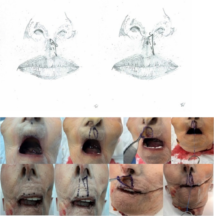 Figure 1
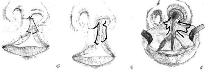 Figure 2
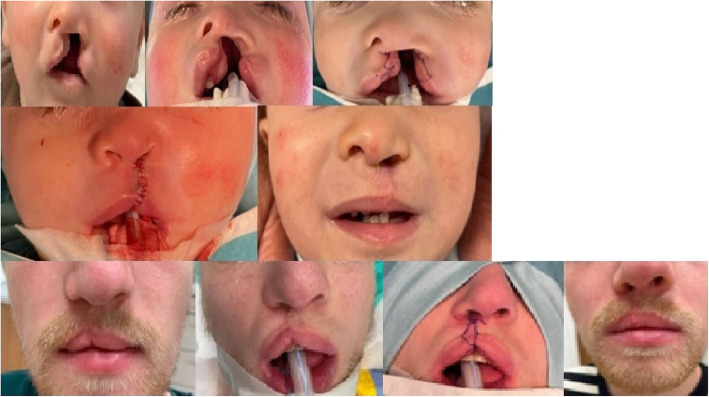 Figure 3
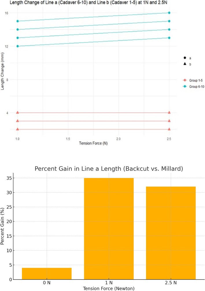 Figure 4
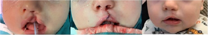 Figure 5
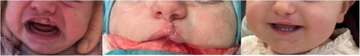 Figure 6
Peer Reviews
No public reviews on file for this paper yet. If you reviewed it on a platform where reviews are public (OpenReview, ICLR, NeurIPS, ICML), you can paste yours below so the community can read it here.
Videos
No videos yet. Explain this paper in a talk, walkthrough, or lecture? Add one.
Taxonomy
TopicsCleft Lip and Palate Research · Reconstructive Surgery and Microvascular Techniques · Craniofacial Disorders and Treatments
Introduction
Long-term results in unilateral cleft lip surgery depend on the visibility of the remaining scar and the scar retraction, which often creates an unpleasant whistling deformity. However, the most important procedure for a pleasing result in cleft lip surgery is a complete mobilization of the orbicularis muscle and its exact adaptation before starting the skin suture. Retraction is the most common issue addressed by different incision patterns for lengthening of native skin at the cleft side of the philtrum. The most commonly used technique of Millard [1], and its derivatives, such as of Mohler [2], create a less appealing scar below the lower nostril due to the c-flap. Millard’s technique, as well as other approaches like Murawski’s [3], where part of the incision line runs below or across the basal central part of the columella, also lead to more visible scarring. Alternative techniques, such as Pfeifer’s, create a straight line with minimal scarring but are more often associated with scar contraction and whistling deformities [4]. The Tennison-Randall technique has a major drawback: disruption of the philtrum by disregarding the aesthetic subunit borders of the upper lip [5]. Similarly, Fisher’s exact subunit technique [6] has disadvantages, including lateral lip tissue scarification [7]. Even in 2024, the search for optimized incision patterns in cleft lip surgery remains ongoing [8]. Dissatisfied with our long-term results using the Millard 2 technique, we sought to develop a technique that combines effective lip lengthening with reduced visible scarring and minimized lip retraction. To interrupt the vertical retraction vector and lengthen the lip, we incorporated the Z-plastic principle at a less conspicuous location. By screening the literature from the past 20 years, we also integrated Fisher’s skin triangle [6, 9], and the transverse mucosal release technique described by Chiang et al. [10] and Baek et al. [11].
Material and methods
Material
After screening the relevant literature, we conducted a cadaver study to gain theoretical insights into the physical properties of skin distraction. No previous study has demonstrated, in a cadaveric model, the extent of lip lengthening achievable with Millard’s technique compared to others. Ten intact adult cadaveric heads, without any signs of trauma, were obtained from the willed donor program at the affiliated medical university cadaver lab. Age and nationality were unknown. Pediatric heads were unavailable; therefore, the effect of aging skin on elasticity could not be measured. When searching actual public databases for studies on biomechanical properties of the skin, we found a recent comprehensive review paper on this topic with the main result that aging skin becomes thinner, stiffer, less tense, and less flexible. Skin tension measured during in vivo uniaxial load and elasticity modules are higher in children than in elderly adults. Mean ultimate skin deformation before bursting is 75% for newborns and 60% for the elderly [12]. From this, we conclude that there is a certain comparability between the skin of old and young people. All specimens were fresh-frozen and defrosted immediately before dissection. In five of the ten specimens, standard Millard advancement-rotation flaps were dissected under loupe magnification on the left side of the philtrum. In the remaining five specimens, back-cut incision flaps were raised as part of our designated Z-back-cut cheiloplasty. We measured the length of the incision lines in Millard’s advancement-rotation flap (see Fig. 1a–c, left) and our back-cut incision (see Fig. 1a–c, right) under varying tension forces (see Fig. 1, below). Line a runs from the center of the lower base of the columella in a straight line to the highest lateral point of the Cupid’s bow. Line b runs from the same starting point to the identical end point, but according to Millard’s incision technique, and line c represents the maximum distance between these two lines of the left part of Fig. 1. On the right side of Fig. 1, line a is identical, while c represents the line from the starting point of line a along the lower base of the columella to its lateral edge with an extension to the highest point of Cupid’s bow, line b. These tension forces were derived from our own clinical experience during unilateral cleft lip repair. The measurements were taken twice per different tensile stress using an elastic stainless-steel ruler with 0.5 mm marking increments. However, measurements were only taken to an accuracy of 1 mm to reduce bias. Cadaver measurements were done exclusively by the corresponding author to reduce inter-observer variability.Fig. 1. Incision lines in our cadaver model; left according to Millard (line b) and right with a straight line with a back-cut (line c and b). Drawing of Millard incision, back-cut incision, and tension force lengthening test. Starting point of line a on the left: Center of the base of the columella. End point of line a on the left: highest lateral point of the cupid’s bow. Line b on the left: identical start and end points. Line c on the left represents the maximum distance between line a and line b. Line a on the right is identical to the left side, starting point of line c is as for line a, but runs along the lower base of the columella to its lateral edge and extends caudally to the end point of line a on the right
As force increased, line a asymptotically approached line b; therefore, no further measurements of line c were conducted in the Millard group. The resulting length of the back-cut technique under tensile stress was calculated as line a, roughly derived from the sum of line b and c. Results were presented in tabular forms (see Tables 1 and 2). Data visualization and statistical analysis were performed using the software package R [13]. We found a lip lengthening effect of the back-cut incision up to 35% compared to the Millard design. Figure 2 supported by the cadaveric findings, we introduced our incision pattern in clinical practice. The Institutional Review Board classification was stated “exempt”, as known surgical techniques were used in combination. Table 1. Lengthening in the craniocaudal direction of the lip with the Millard techniqueTechnique: MillardTension force in Newton NLine012,55SexTeethCadaver 1 line length in mma22303232FemalenoCadaver 1 line length in mmb28323232Cadaver 1 line length in mmc6Cadaver 2 line length in mma19232424MaleYesCadaver 2 line length in mmb22252525Cadaver 2 line length in mmc6Cadaver 3 line length in mma16212222MaleYesCadaver 3 line length in mmb18222222Cadaver 3 line length in mmc5Cadaver 4 line length in mma18252626FemaleNoCadaver 4 line length in mmb23262626Cadaver 4 line length in mmc6Cadaver 5 line length in mma21252626FemaleNoCadaver 5 line length in mmb25272727Cadaver 5 line length in mmc6Mean line a19.224.82626Mean line b23.226.426.426.4SD line a2.393.353.743.74SD line b3.73.653.653.65Lengthening in the craniocaudal direction of the lip with the Millard technique with mean and SD for line a and bTable 2Lengthening in the craniocaudal direction of the upper lip with the back-cut techniqueTechnique: backcut-cheiloplastyTension force in Newton NLine012,55SexTeethCadaver 6 line length in mma17303131malenoCadaver 6 line length in mmb17212222Cadaver 6 line length in mmc7Cadaver 7 line length in mma21333434femalenoCadaver 7 line length in mmb22242525Cadaver 7 line length in mmc8Cadaver 8 line length in mma20333434MaleNoCadaver 8 line length in mmb22232424Cadaver 8 line length in mmc7Cadaver 9 line length in mma20353636FemaleYesCadaver 9 line length in mmb22242525Cadaver 9 line length in mmc8Cadaver 10 line length in mma22363737MaleYesCadaver 10 line length in mmb24262727Cadaver 10 line length in mmc8Mean line a2033.434.434.4Mean line b21.423.624.624.6SD line a1.872.32.32.3SD line b2.611.821.821.82Lengthening in the craniocaudal direction of the upper lip with the back-cut technique with mean and SD for line a and b
Patients
Patients with partial or complete unilateral cleft lip or cleft lip/alveolus/palate were selected. Surgery was performed by the first author using the technique described below. Individuals with complete cleft lip and palate were treated similarly to those with unilateral cleft lip. Postoperative follow-up adhered to our cleft team protocol. However, due to the COVID-19 pandemic, delays occurred in treatment and follow-up beginning in January 2020 (patients characteristics see below).
GenderDate of birthDate of operationAge at the operation (month)Type of cleftFollow-up on cleft teamComplications MaleMay 2023March 202410Complete lip/bone/palate leftApril 2025Complete dehiscence of the soft palate and partial dehiscence of the lip caused by an infectionMaleDecember 2022August 20238Partial lip/bone leftSeptember 2023NoneFemaleOctober 2022August 202310Partial lip/bone leftDecember 2024NoneFemaleFebruary 2023November 20238Complete lip/bone leftJanuary 2024NoneMaleMarch 2000February 202423 yearsComplete lip/bone/palate rightNovember 2024NoneFemaleJanuary 2022February 202313Complete lip/bone/palate rightApril 2024Complete dehiscence of the soft palate and lost to follow-up since April 2024FemaleMay 2022May 202311Complete lip/bone/palate leftNovember 2024Postop. airway obstruction caused by a combined Furlow plastyMaleAugust 2020March 202111Partial lip/bone leftFebruary 2023NoneFemaleSeptember 2019January 20203Complete lip/bone/palate left with SimonardDecember 2024Mild hypertrophic scarring of the lipFemaleSeptember 2019October 202013Complete lip/bone/palate left by microdeletionJanuary 2025NoneFemaleDecember 2021August 20227Partial lip/bone leftJune 2024NoneFemaleFebruary 2020February 202111Complete lip/bone with ocular hypertelorismAugust 2023Hypertrophic scarring as a consequence of a postoperative wound infection of the lip with sec. corrections, lost to follow-up since October 2023MaleMay 2020November 20245Partial lip leftNovember 2022NoneMaleNovember 2019November 202412Partial lip/bone rightFebruary 2025NoneMaleNovember 2003January 202420 yearsComplete lip/bone rightJanuary 2024Lost to follow-up since January 2024MaleMarch 2024January 202510Bilateral complete lip/bone/palateFebruary 2025NoneMaleJuly 2024Februayr 20256Complete lip rightMarch 2025None
Drawings and surgical technique
The incision design is illustrated in Fig. 2. Initial steps include marking of Cupid’s bow, the subnasal, the lateral base of the columella on the cleft side, and an additional perpendicular marking 2 mm cranial to the cleft-side peak of Cupid’s bow in the white roll of the vermilion border under loupe magnification. From this point, a line is drawn just outside the vermilion border toward the nasal sill, with a back cut extending to the marked lateral columella base. From the Noordhoff point (the point along the lateral lip where the vermillion height is at its greatest and the white roll is well formed but becomes less distinct toward the cleft as the progressive deficiency of the orbicularis oris muscle), the incision is extended outside the vermilion border towards the cleft sill, incorporating a small Fisher’s triangle (as small triangular pennant skin flap above of Noordhoff point) cranially in the lateral white roll. A medial triangular skin flap is added at the nostril base. The dry/wet line of the vermilion is marked bilaterally. Small distal triangular flaps of the dry vermilion skin are created for additional lip volume. For complete clefts, two mucosal vermilion flaps are dissected to close the nasal floor. In incomplete clefts, they can be discarded. The orbicularis oris muscle and its subdivisions, such as the depressor septum nasalis muscle, are released from the base of the columella, nasal ala, and the alveolar cleft of the maxillary bony segment with a single or double mucosal incision at the oral vestibular sulcus. The vestibular mucosa is sutured with interrupted Vicryl 4.0 sutures, followed by cranial-to-caudal adaptation of the orbicularis oris muscle with PDS 4.0 and Vicryl 4.0 sutures. Skin closure begins at the vermilion border using Monocryl 6.0. The contralateral relaxing back-cut incision is positioned precisely in the sulcus of the contralateral white roll for Fisher’s pennant flap placement. The small flap is trimmed and sutured into place, followed by nostril closure. The triangular flaps of the Z-plasty at the columella base are adjusted to size before final closure. Excess mucosa is resected before placing steristrips across the suture line. In complete cleft lip and palate cases, primary rhinoplasty following McComb’s method (with blunt mobilization of the nasal skin around the lower latera) is performed, with a silicone nostril retainer placed for 6 to 8 weeks. The technique may also be suitable for secondary lip revision in adults (see Figs. 3, 4, 5, and 6). To date, a total of 17 patients with a partial or complete cleft lip have been operated on using the technique described here in the last 4 years. So far, one secondary revision has been performed; according to our cleft team protocol, these are in most cases-only performed in adolescence, sometimes in combination with rhinoplasty.
Fig. 2. Incision pattern on a left partial cleft lip, complete cleft lip, and intraoral lines at the intraoral lip sulcus
Fig. 3. Above: complete cleft lip and palate left at 4 months of age with presurgical taping, intraoperative drawings with immediate result and 6 months postoperative at the age of 1.5 years with a postoperative wound infection caused by continuously nostril retainer manipulation by the child and partial dehiscence at the alar base with scar contraction. Below: Z-back cut cheiloplasty at the age of 24 with preoperative, intraoperative, and postoperative photos at 6 months including the drawing pattern Fig. 4. No increase in the length chance of line a in Millard’s incision line. Increase of lengthening of line a in the group of back-cut incision compared to line b in Millard design with 1N and 2.5N tension force. No further lengthening of both lines with 5N. With increasing tension, line a approximates line b in Millard’s technique, while lines b+c approximates line a in the back-cut technique. Lower Part: Percentage gain in length between the two incision patterns Fig. 5. Operated upper lip at the age of 9 months, intraoperative result and 7 weeks postoperatively with our technique by partial cleft of the lip and alveolus Fig. 6. Partial cleft of the left upper lip with closure by Z-back cut-cheiloplasty preoperatively at 2 weeks of age, direct intraoperatively at the age of 10 months and at 6 weeks postoperatively
Our tension force studies demonstrated insufficient lengthening of Millard’s advancement-rotation flap compared to our back-cut incision (see Fig. 4). Between 2.5 N and 5 N tension, no further lengthening occurred; thus, statistical analysis at 5 N was omitted. Comparisons between the two incision patterns at 1 N and 2.5 N showed no significant elongation with Millard’s incision, whereas the back-cut technique produced a significant increase. The results were highly statistically significant (p < 0.001, Student’s t-test). This suggests that Millard’s technique does not appear to achieve the expected skin lengthening via rotation and advancement. In contrast, an incision along the philtral column with a back-cut at the cleft-side columella base, as part of a Z-plasty, significantly enhances lengthening. Furthermore, the resulting straight line reassembles the lateral border of the philtrum more accurately. The resulting defect of the back-cut incision at the lateral base of the columella can be filled up with the cutaneous triangle of the contralateral side of the cleft as part of a Z-plasty.
Discussion
Our study correlates incision-line lengthening in unilateral cleft lip repair with applied tension forces. The primary and most important limitation is the use of adult cadaveric skin. However, we believe the lengthening effect is largely independent of age, as it is based on the geometric principle of Z-plasty [14, 15]. The data indicate that the distance between the columella base and Cupid’s bow lateral border depends on the incision pattern. This tension line forms the future philtral column on the cleft side post-surgery. To maximize lengthening, we recommend combining Z-plasty with Fisher’s pennant flap and relaxing incisions at the gingival sulcus to prevent upper lip under-projection. We also use vermilion mucosal flaps in complete cleft lip and palates to reconstruct the nasal floor and to augment shortness of mucosal lining at the site of the cleft sulcus. As known in Furlow’s palate closure technique or constriction ring syndromes, the Z-plasty principle adds lengthening and less chance for retraction in the long run. The meticulous adapting of all parts—but especially the caudal part—of the upper orbicularis oris muscle seems the most eminent step in cleft lip closure to prevent vermilion notching or even whistling deformity [16] in combination with adequate lengthening of the inner mucosal lining of the cleft repair. The resulting straight line reassembles the lateral border of the philtrum more accurately. The resulting defect of the back-cut incision at the lateral base of the columella can be filled up with the cutaneous triangle of the contralateral side of the cleft as part of a Z-plasty. Furthermore, the central base of the columella stays untouched and free of visible scars. Over the past 4 years, we have performed our technique on 17 patients. Although long-term results remain pending, we hope to expect less secondary lip revisions due to reduced vertical scar contracture. We are well aware that cadaveric skin behaves differently from pediatric skin and therefore it is difficult to draw direct conclusions from our cadaver study. Follow-up of these children is a prerequisite to estimate the value of our developed technique. We also believe our technique is well-suited for bilateral cleft lip repair due to its additional philtrum lengthening capabilities. This novel incision pattern has not been previously published and represents a valuable addition to existing techniques by incorporating the best elements from previous approaches.
The reference list from the paper itself. Each links out to its DOI / PubMed record.
- 1Team RC. R Core Team. R: A language and Invironment for statistical Computing. R Foundation for Statistical Computing, Vienna, Austria. https://www.R-project.org/. 2022.