The CT target sign as a criterion for the differential diagnosis between tuberculosis and organizing pneumonia
Elcio Bakowski, Gláucia Zanetti, Edson Marchiori

Abstract
Genes, proteins, chemicals, diseases, species, mutations and cell lines named across the full text — each resolved to its canonical identifier and authoritative record.
Click any figure to enlarge with its caption.
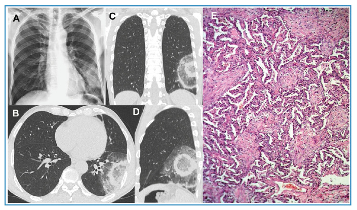 Figure 1
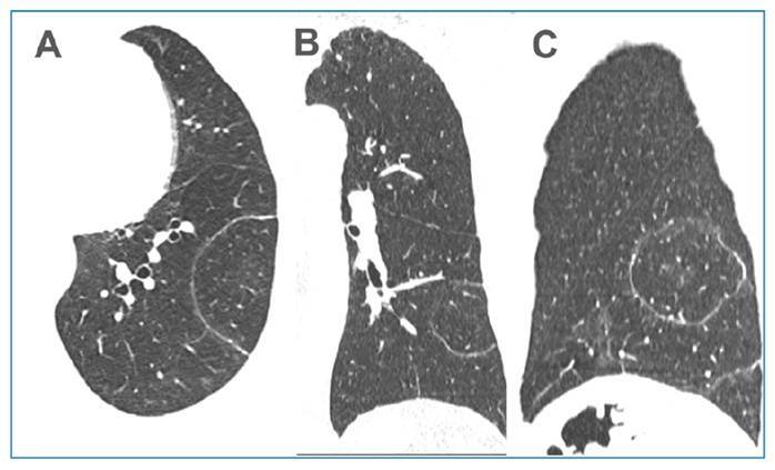 Figure 2
Peer Reviews
No public reviews on file for this paper yet. If you reviewed it on a platform where reviews are public (OpenReview, ICLR, NeurIPS, ICML), you can paste yours below so the community can read it here.
Videos
No videos yet. Explain this paper in a talk, walkthrough, or lecture? Add one.
Taxonomy
TopicsTuberculosis Research and Epidemiology · Infectious Diseases and Tuberculosis · Diagnosis and treatment of tuberculosis
A 61-year-old male patient developed fever, dry cough, myalgia, headache, and arthralgia and was treated with prednisone and symptom-based medications. The decision to initiate corticosteroid therapy instead of antibiotics was based on the atypical clinical and laboratory features of a bacterial infection, which led us to consider it an inflammatory or autoimmune disease. Five days later, the patient’s condition worsened and was hospitalized. The patient denied any other signs or symptoms. Complementary examinations including a respiratory viral panel yielded no significant findings. Chest radiography revealed inhomogeneous opacities in the left lower lobe of the lung (Figure 1A). Chest CT findings were compatible with the target sign, which was interpreted as organizing pneumonia (Figure 1B-D). The patient underwent bronchoscopy, with bronchoalveolar lavage and biopsy. Real-time PCR (GeneXpert) was performed on bronchoalveolar lavage samples, and Mycobacterium tuberculosis was detected. A regimen consisting of RIPE (rifampin, isoniazid, pyrazinamide, and ethambutol) was initiated. Five days later, transbronchial biopsy showed a histological pattern of organizing pneumonia (Figure 1E), confirming the tomographic diagnosis. The RIPE regimen was considered a false-positive result and suspended based on the tomographic findings of the target sign and the results of the transbronchial biopsy; then, prednisone was started. The patient was discharged from hospital in good clinical condition. Three weeks after the initial examination, a control CT scan showed a marked reduction in the lesions (Figure 2).
FIGURE 1: (A) Chest radiograph showing inhomogeneous opacities in the left lower lobe of the lung. Non-enhanced chest CT images with axial (B), coronal (C) and sagittal (D) reconstruction demonstrate a focal core ground-glass opacity and two ring-like opacities immediately surrounding it, with the appearance of a shooting target (the CT target sign). A photomicrograph of the lung biopsy (E) shows the involvement of airspaces by polypoid fibroblastic foci distributed within terminal bronchioles, alveolar ducts, and alveoli (hematoxylin and eosin stain, ×40).
FIGURE 2:Non-enhanced chest CT images acquired three weeks after the first examination with axial (A), coronal (B) and sagittal (C) reconstruction show marked resorption of the opacities, with only a dense halo persisting, involving the normal lung parenchyma.
The target sign consists of a central nodular opacity with variable densities (soft tissue or ground glass) surrounded by a less dense rim of either normal parenchyma or ground glass opacity. This is further encircled by a denser peripheral rim of ground glass or soft tissue-type density. In some cases, multiple concentric ring-like opacities may be observed1 ^-^ 3. In this instance, recognizing the target sign strongly suggests a diagnosis of organizing pneumonia3 ^-^ 5. To our knowledge, this sign has not been documented in patients with pulmonary tuberculosis.
The reference list from the paper itself. Each links out to its DOI / PubMed record.
- 1Marchiori E Penha D Nobre LF Hochhegger B Zanetti G Differences and Similarities Between the Chest CT Target Sign and the Reversed Halo Sign in Patients with COVID-19 Pneumonia Korean J Radiol 202122467267610.3348/kjr.2020.115033660464 PMC 8005353 · doi ↗ · pubmed ↗
- 2Marchiori E Marques Silva JA Amorim VB Zanetti G Is the CT target sign specific to COVID-19 pneumonia?J Bras Pneumol 2020466 e 2020054110.36416/1806-3756/e 20200541 · doi ↗
- 3Müller CIS Müller NL Chest CT target sign in a couple with COVID-19 pneumonia Radiol Bras 202053425225410.1590/0100-3984.2020.008932904794 PMC 7458560 · doi ↗ · pubmed ↗
- 4Jafari R Jonaidi-Jafari N Maghsoudi H Dehghanpoor F Schoepf UJ Ulversoy KA Saburi A "Pulmonary target sign" as a diagnostic feature in chest computed tomography of COVID-19World J Radiol 2807202113723324210.4329/wjr.v 13.i 7.23334367510 PMC 8326149 · doi ↗ · pubmed ↗
- 5Mehrabi Nejad MM Salehi M Azadbakht J Jahani Z Veisi P Sedighi N Salahshour S Is target sign (bull's eye appearance) associated with adverse outcomes in COVID-19 patients? A case series and literature review Caspian J Intern Med 202213 Suppl 327027610.22088/cjim.13.0.27035872681 PMC 9272949 · doi ↗ · pubmed ↗