Dynamic PET imaging in patients with unilateral carotid occlusion shows lateralized cerebral hypoperfusion, but no amyloid binding
Naomi LP Starmans, Anna E Leeuwis, Edwin Bennink, Sebastiaan L Meyer Viol, Sandeep SV Golla, Jan Willem Dankbaar, Esther E Bron, Geert Jan Biessels, L Jaap Kappelle, Wiesje M van der Flier, Nelleke Tolboom

TL;DR
This study found that patients with a blocked carotid artery have reduced blood flow in the affected brain hemisphere, but no increase in amyloid buildup or cognitive issues linked to it.
Contribution
The study is the first to show that unilateral carotid occlusion causes lateralized hypoperfusion without increased ipsilateral amyloid-β accumulation.
Findings
Ipsilateral cerebral perfusion was significantly lower compared to the contralateral hemisphere.
No significant difference in amyloid-β binding was found between hemispheres.
Neither perfusion nor amyloid-β levels were associated with cognitive performance.
Abstract
Carotid occlusive disease is a risk factor for cognitive decline. A possible underlying etiology is that hemodynamic impairment results in decreased cerebral perfusion, exacerbated amyloid-β accumulation (Aβ) and poorer cognitive performance. We aimed to determine whether patients with unilateral internal carotid artery (ICA) occlusion have less cerebral perfusion and more Aβ in the ipsilateral than in the contralateral hemisphere, and whether perfusion and Aβ are associated with cognitive functioning. We included 20 patients (age 67.2 ± 7.0 years, 8 females, MMSE 29 [27–29]) with unilateral ICA occlusion, which underwent neuropsychological assessment and dynamic 18F-Florbetaben positron emission tomography (PET). Global and regional relative perfusion (R1) and binding potential (BPND) were obtained from the PET-images using a simplified reference tissue model. We performed Wilcoxon…
Genes, proteins, chemicals, diseases, species, mutations and cell lines named across the full text — each resolved to its canonical identifier and authoritative record.
Click any figure to enlarge with its caption.
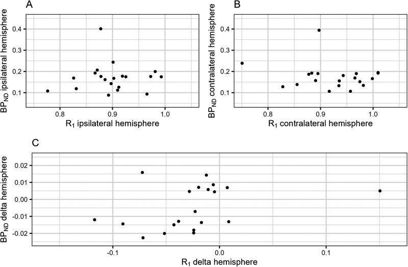 Figure 1
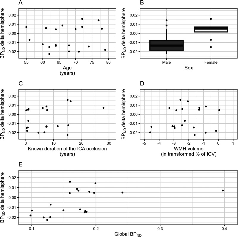 Figure 2
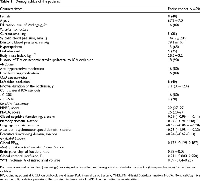 Figure 3
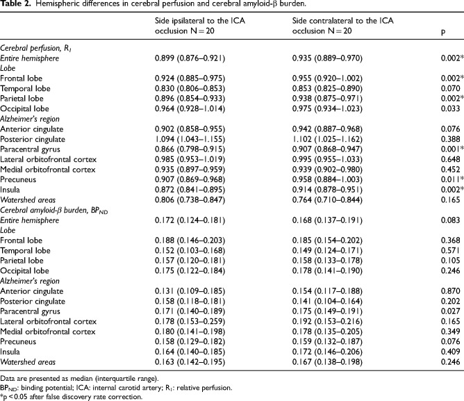 Figure 4
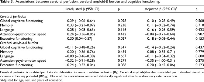 Figure 5
Peer Reviews
No public reviews on file for this paper yet. If you reviewed it on a platform where reviews are public (OpenReview, ICLR, NeurIPS, ICML), you can paste yours below so the community can read it here.
Videos
No videos yet. Explain this paper in a talk, walkthrough, or lecture? Add one.
Taxonomy
TopicsCerebrovascular and Carotid Artery Diseases · Neurological Disease Mechanisms and Treatments · Dementia and Cognitive Impairment Research