Evaluation of Stress Distribution and Displacement in the Affected Periodontium of Mandibular Incisors Using Utility Intrusion and Reverse Curve of Spee Arch Wires: A Finite Element Analysis
Vazrala Vamsi Krishna Reddy, Ghanta Sunil, Soma Balaji, Soorabathula Sonika Mani kiran, Konni Prudhvi, RSVM Raghu Ram

TL;DR
This study uses computer modeling to compare two orthodontic wires in managing stress and tooth movement in cases of bone loss around lower front teeth.
Contribution
The study introduces a finite element analysis comparing utility intrusion and reverse curve of Spee arch wires in varying levels of bone loss.
Findings
Higher bone loss increases stress and decreases displacement, leading to labial tipping.
Utility intrusion arch wire shows better torque control and less labial tipping than the reverse curve of Spee wire.
Stress distribution is consistently higher with the utility intrusion arch wire across all bone loss levels.
Abstract
Objective: This study aims to evaluate stress distribution and displacement in the affected periodontium of mandibular incisors with utility intrusion arch wire and reverse curve of Spee (RCS) arch wire using finite element analysis (FEA). Materials and methods: A 3D finite element model (FEM) of the mandibular arch was created, simulating mandibular incisors with three variations of bone loss (0%, 33%, and 66%) and 0.022” x 0.028” slot MacLaughlin, Bennett, and Trevisi prescription brackets. Utility intrusion arch wire and RCS arch wire were modelled using 0.017” x 0.025 stainless steel and subjected to activation. Stress distribution and displacement were calculated and analyzed. Results: Comparison between utility intrusion arch wire and RCS arch wire, using FEM for mandibular incisors with varying bone loss (0%, 33%, and 66%), revealed alteration in the stress distribution and…
Genes, proteins, chemicals, diseases, species, mutations and cell lines named across the full text — each resolved to its canonical identifier and authoritative record.
Click any figure to enlarge with its caption.
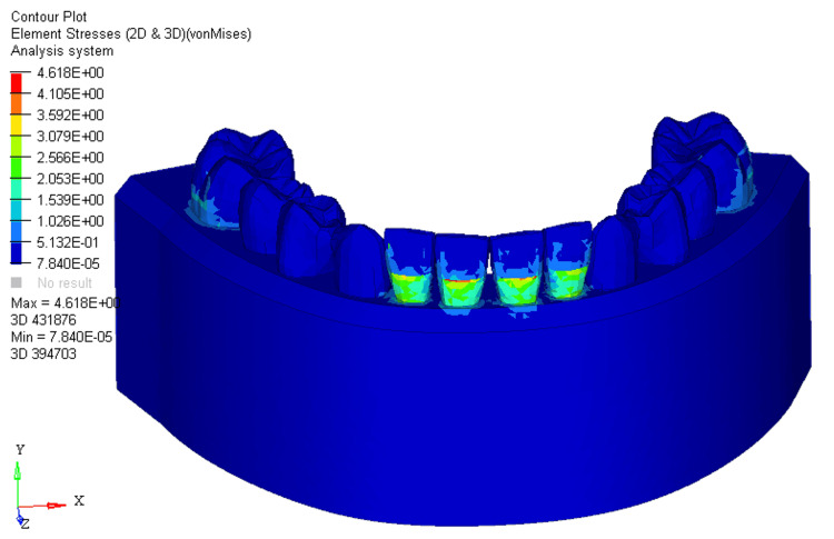 Figure 1
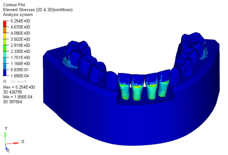 Figure 2
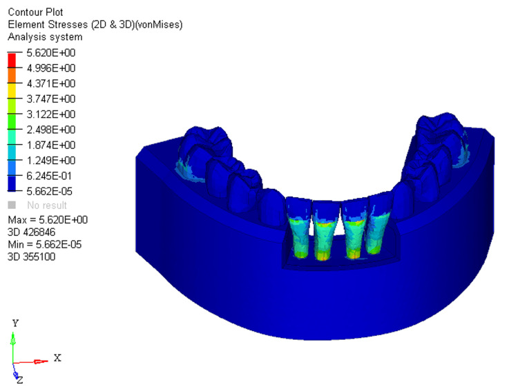 Figure 3
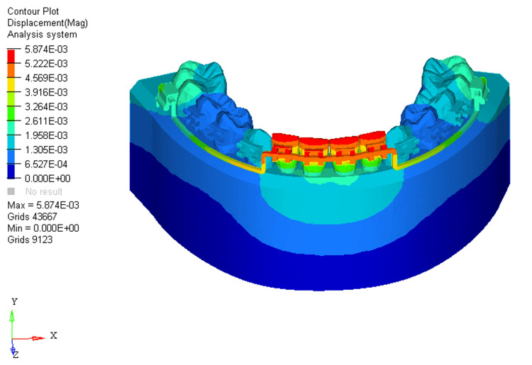 Figure 4
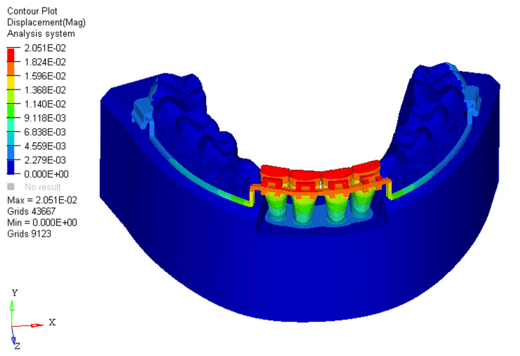 Figure 5
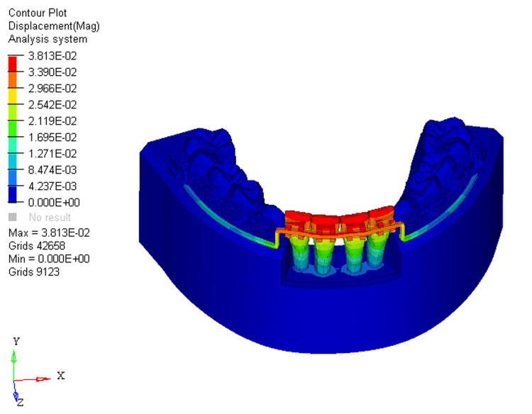 Figure 6
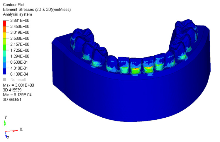 Figure 7
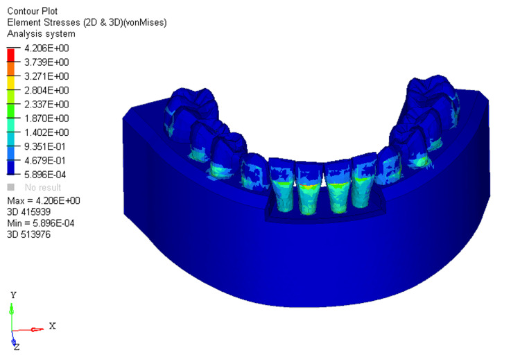 Figure 8
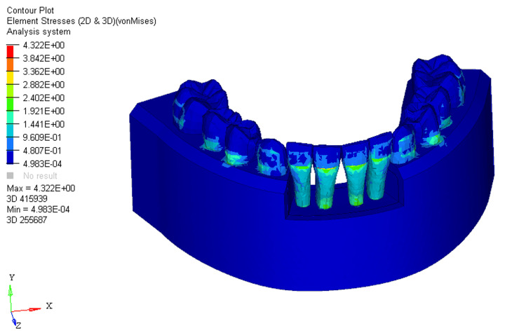 Figure 9
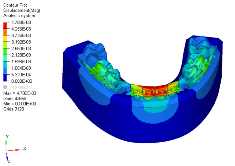 Figure 10
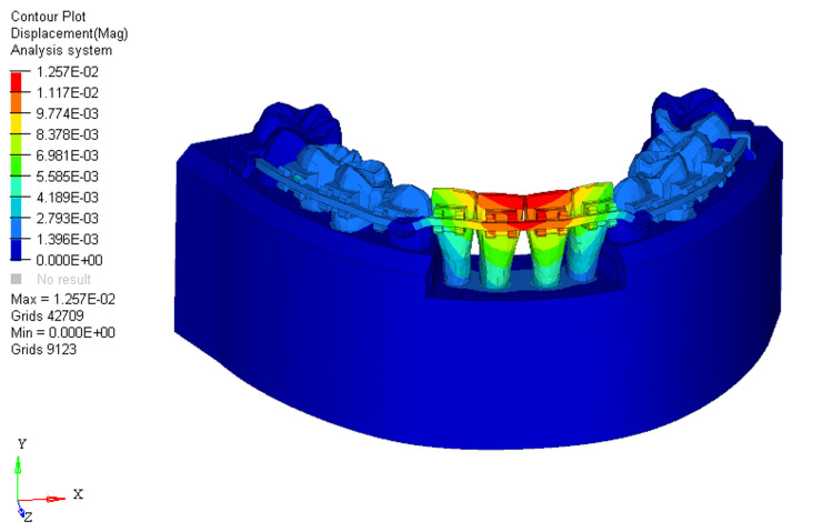 Figure 11
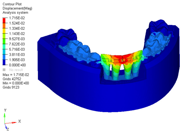 Figure 12
Peer Reviews
No public reviews on file for this paper yet. If you reviewed it on a platform where reviews are public (OpenReview, ICLR, NeurIPS, ICML), you can paste yours below so the community can read it here.
Videos
No videos yet. Explain this paper in a talk, walkthrough, or lecture? Add one.
Taxonomy
TopicsOrthodontics and Dentofacial Orthopedics · Dental Radiography and Imaging · dental development and anomalies