Diagnostic Utility of Diffusion-Weighted MRI and Apparent Diffusion Coefficient Values in Differentiating Metastatic From Non-metastatic Lymph Nodes in Cervical Carcinoma
Vamsi Venkat, Anil K Sakalecha, Anees Dudekula, Mahima Kale R, Guru Yogendra Muthyal, Kalyani R

TL;DR
This study shows that diffusion-weighted MRI and ADC values can effectively distinguish between cancerous and non-cancerous lymph nodes in cervical cancer patients.
Contribution
The study demonstrates that ADC values can reliably detect metastatic lymph nodes in cervical cancer without histopathological confirmation.
Findings
Metastatic lymph nodes had significantly lower ADC values (~0.90 × 10⁻³ mm²/s) compared to non-metastatic nodes (~1.30 × 10⁻³ mm²/s).
An ADC threshold of 1.0 × 10⁻³ mm²/s achieved 93% sensitivity and 100% specificity for detecting metastatic nodes.
DWI and ADC mapping outperformed traditional size-based criteria for identifying metastatic lymph nodes.
Abstract
Background: In cervical carcinoma, lymph node involvement is a key indicator of disease progression and a critical prognostic factor that influences staging and treatment. Accurate identification of metastatic lymph nodes is essential for optimal management. Diffusion-weighted MRI combined with apparent diffusion coefficient (ADC) mapping provides a non-invasive approach for evaluating tissue cellularity and may enhance the identification of metastatic lymph nodes, surpassing traditional size-based criteria. Purpose: The purpose of the study is to assess the effectiveness of diffusion-weighted imaging (DWI) and ADC values in differentiating between metastatic and non-metastatic lymph nodes in patients with cervical cancer. The study focuses on imaging findings without histopathological confirmation, aiming to determine whether ADC measurements can reliably indicate nodal metastasis.…
Genes, proteins, chemicals, diseases, species, mutations and cell lines named across the full text — each resolved to its canonical identifier and authoritative record.
Click any figure to enlarge with its caption.
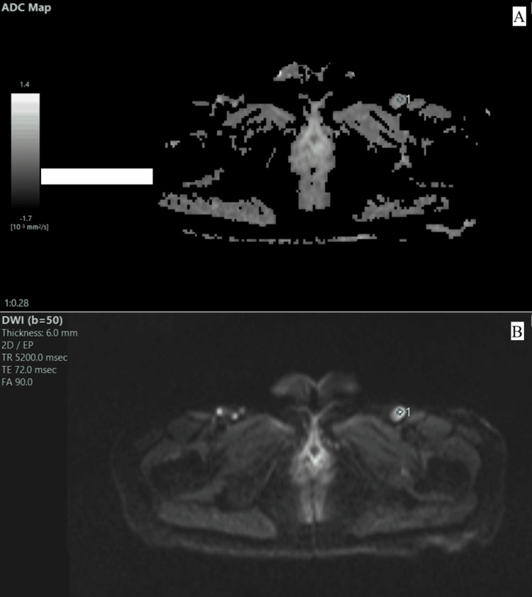 Figure 1
Peer Reviews
No public reviews on file for this paper yet. If you reviewed it on a platform where reviews are public (OpenReview, ICLR, NeurIPS, ICML), you can paste yours below so the community can read it here.
Videos
No videos yet. Explain this paper in a talk, walkthrough, or lecture? Add one.
Taxonomy
TopicsMRI in cancer diagnosis · Radiomics and Machine Learning in Medical Imaging · Endometrial and Cervical Cancer Treatments