A novel peroral cholangioscopy of gallbladder carcinosarcoma: a case report
Takumi Onoyama, Taro Yamashita, Takuya Shimosaka, Yuri Sakamoto, Noriyuki Suto, Tsuyoshi Mikamo, Hajime Isomoto

Abstract
Genes, proteins, chemicals, diseases, species, mutations and cell lines named across the full text — each resolved to its canonical identifier and authoritative record.
Click any figure to enlarge with its caption.
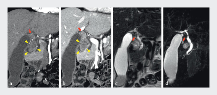 Fig. 1
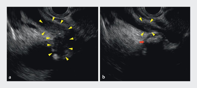 Fig. 2
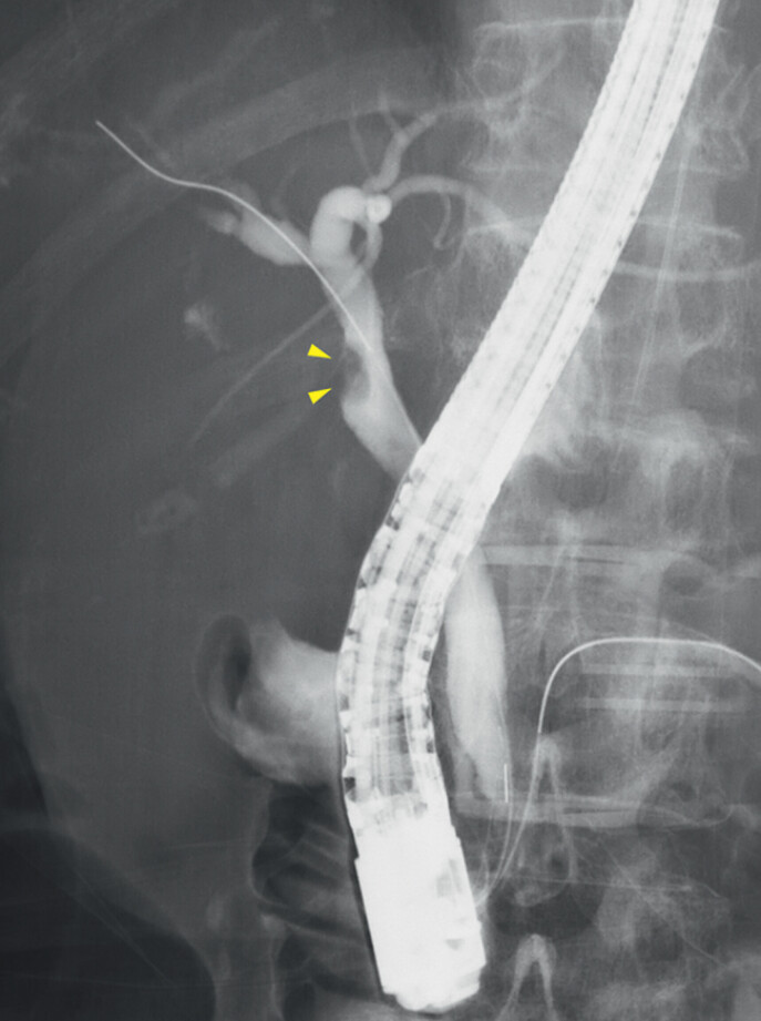 Fig. 3
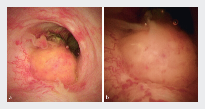 Fig. 4
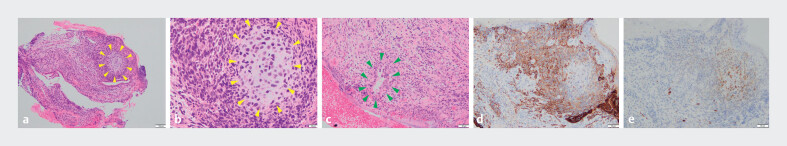 Fig. 5
Peer Reviews
No public reviews on file for this paper yet. If you reviewed it on a platform where reviews are public (OpenReview, ICLR, NeurIPS, ICML), you can paste yours below so the community can read it here.
Videos
No videos yet. Explain this paper in a talk, walkthrough, or lecture? Add one.
Taxonomy
TopicsCholangiocarcinoma and Gallbladder Cancer Studies · Cancer Diagnosis and Treatment · Metastasis and carcinoma case studies
Gallbladder carcinosarcoma (GBCS) is extremely rare, accounting for less than 1 % of malignant primary gallbladder tumors with poor prognosis 1 2 3 4 . Due to its rarity, the literature on GBCS is limited, with only approximately 100 cases reported 5 . We herein present a case of GBCS in which the tumor was visualized directly using a novel peroral cholangioscope (eyeMax; Micro-Tech, Nanjing, China).
A 74-year-old female visited a medical institution due to loss of appetite, where a gallbladder mass was detected, and she was admitted to our hospital. Contrast-enhanced abdominal computed tomography showed a mass from the gallbladder neck to the common bile duct (CBD). An enlarged Lymph node with contrast enhancement was observed between the extrahepatic bile duct and gallbladder ( Fig. 1 a, b ). The patient also underwent magnetic resonance cholangiopancreatography, which demonstrated a filling defect in the CBD, near the junction of the cystic duct ( Fig. 1 c, d ). Endoscopic ultrasonography revealed a hypoechoic lesion in this region ( Fig. 2 a, b ). Endoscopic retrograde cholangiopancreatography was performed to confirm the diagnosis of the biliary lesion and revealed a filling defect at the junction of the cystic duct ( Fig. 3 ). Peroral cholangioscopy (POCS) revealed an irregular subepithelial-like lesion with irregularly dilated vessels at the junction of the cystic duct, which was suspected to be a malignant tumor ( Fig. 4 a, b and Video 1 ). The histopathological findings of the forceps biopsies from the lesion revealed the diagnosis of carcinosarcoma, which consists of adenocarcinoma, sarcomatous cells, and chondroid matrix ( Fig. 5 ). POCS-guided mapping biopsy also showed that atypical epithelial cells exist in the confluence of intrahepatic bile ducts. The patient preferred best supportive care over aggressive treatment. To the best of our knowledge, this is the first report of the visualization of GBCS using a novel POCS.
Computed tomography image and magnetic resonance cholangiopancreatography. a, b A mass from the gallbladder neck to the extrahepatic bile duct was identified (arrows). Enlarged Lymph node with contrast enhancement was observed between the extrahepatic bile duct and gallbladder (arrowheads). c, d Filling defect in the biliary system, from the gallbladder neck to the junction of the cystic duct (arrows). There was no dilation in the common hepatic duct.
Endoscopic ultrasonography. a, b There was an irregular hypoechoic lesion from the gallbladder neck to the extrahepatic bile duct (arrowheads). A gallbladder stone exists near the lesion (arrow).
Endoscopic retrograde cholangiopancreatography showing. A filling defect is observed in the common bile duct near the junction of the cystic duct (arrowheads).
Peroral cholangioscopy. a, b There was a subepithelial-like lesion with irregularly dilated vessels at the junction of the cystic duct.
The video shows a case of gallbladder carcinosarcoma in which the tumor was visualized directly using a peroral cholangioscope. It seemed a subepithelial-like lesion with irregularly dilated vessels.Video 1
Hematoxylin and eosin staining images of the forceps biopsy sample. a, b Chromatin-rich short spindle cells are proliferating diffusely within the stroma. The chondroid matrix was also observed in the stroma (yellow arrowheads). c Atypical cells with irregular nuclei and disrupted polarity were proliferating, forming glandular ducts (green arrowheads). The above findings suggested a mixture of adenocarcinoma, sarcomatous cells, and chondroid matrix, and it was diagnosed as carcinosarcoma. d Adenocarcinoma and sarcomatous components were positive with immunohistochemical staining for pan-cytokeratin (CK AE1/AE3). e The chondroid matrix was immunohistochemically positive for S100.
Endoscopy_UCTN_Code_CCL_1AZ_2AC
The reference list from the paper itself. Each links out to its DOI / PubMed record.
- 1Cao R Jiang H Zhang Y Comparison of carcinosarcoma and adenocarcinoma of the gallbladder: a study based on SEER population for propensity matching and nomogram analysis J Cancer Res Clin Oncol 2023149139851399310.1007/s 00432-023-05220-037543541 PMC 11796759 · doi ↗ · pubmed ↗
- 2Teng TZJ Chua BQY Shelat VG Carcinosarcoma of gallbladder: A world review World J Clin Oncol 2021121244126310.5306/wjco.v 12.i 12.124435070742 PMC 8716988 · doi ↗ · pubmed ↗
- 3Okabayashi T Shima Y Iwata J Surgical outcomes for 131 cases of carcinosarcoma of the hepatobiliary tract J Gastroenterol 20144998299110.1007/s 00535-013-0882-224162331 · doi ↗ · pubmed ↗
- 4Okabayashi T Sun ZL Montgomery RA Surgical outcome of carcinosarcoma of the gall bladder: A review World J Gastroenterol 2009154877488219842216 10.3748/wjg.15.4877 PMC 2764963 · doi ↗ · pubmed ↗
- 5Mansour S Derkach E Abergil V Carcinosarcoma of the Gallbladder: A Rare Tumor World J Oncol 20221310310610.14740/wjon 149535837320 PMC 9239502 · doi ↗ · pubmed ↗