Expanded range of Haemagogus leucocelaenus in yellow fever hotspots: new findings from Santa Catarina State, southern Brazil
Sabrina Fernandes Cardoso, Iara Carolini Pinheiro, Larissa Akemi Oliveira Kikuti, Andre Akira Gonzaga Yoshikawa, André Nóbrega Pitaluga, Luísa Damazio P Rona

TL;DR
This study reports new locations of a yellow fever-carrying mosquito in southern Brazil and confirms its identity using DNA analysis.
Contribution
The first confirmed records of Haemagogus leucocelaenus in Santa Catarina State, Brazil, using molecular and morphological methods.
Findings
Haemagogus leucocelaenus was newly recorded in five municipalities in southern Santa Catarina.
Molecular identification confirmed all specimens as Hg. leucocelaenus despite morphological variations.
DNA barcoding provides a reliable method for species identification in this region.
Abstract
The Haemagogus genus includes nine mosquito species reported in Brazil, each with distinct distribution patterns. Haemagogus leucocelaenus, a major yellow fever vector, is widely distributed throughout the country, while Haemagogus leucophoebus, a morphologically similar species, has only been identified in Acre State. This study evaluated the presence of Haemagogus species in southern Brazil by comparing their morphological and molecular characteristics. Mosquitoes were collected from five municipalities in southern Santa Catarina State, Brazil. Each specimen was identified morphologically and photographed. Genomic DNA was extracted, and a Cytochrome C Oxidase Subunit I (COI) gene fragment was amplified using polymerase chain reaction (PCR). The positive amplicons were sequenced for molecular identification. New records of Hg. leucocelaenus were found in Santa Rosa de Lima, Rio…
Genes, proteins, chemicals, diseases, species, mutations and cell lines named across the full text — each resolved to its canonical identifier and authoritative record.
Click any figure to enlarge with its caption.
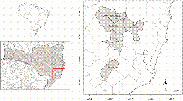 Figure 1
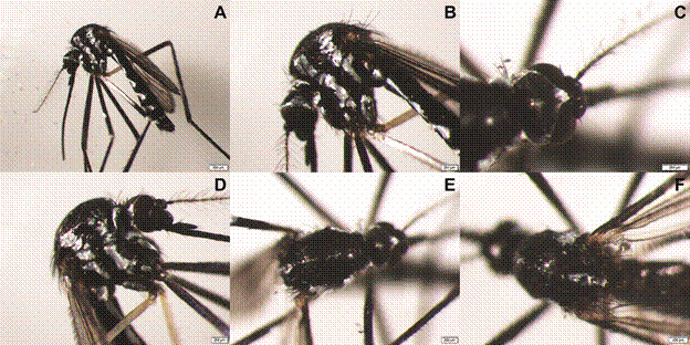 Figure 2
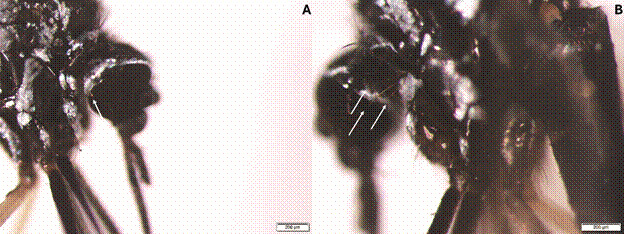 Figure 3
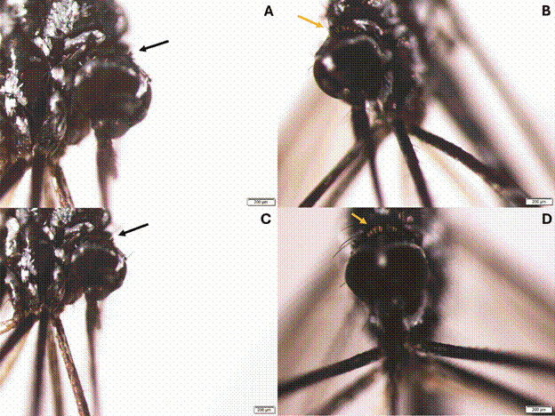 Figure 4
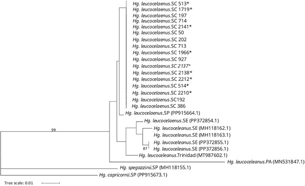 Figure 5
Peer Reviews
No public reviews on file for this paper yet. If you reviewed it on a platform where reviews are public (OpenReview, ICLR, NeurIPS, ICML), you can paste yours below so the community can read it here.
Videos
No videos yet. Explain this paper in a talk, walkthrough, or lecture? Add one.
Taxonomy
TopicsMosquito-borne diseases and control · Vector-borne infectious diseases · Parasite Biology and Host Interactions
Yellow fever (YF) is a non-contagious disease caused by the YF virus (YFV), which belongs to the species Orthoflavivirus flavi 1 ^,^ 2 within the Orthoflavivirus genus of the Flaviviridae family.2 In Brazil, YF is transmitted through two cycles: (i) the sylvatic (or jungle) cycle, involving non-human primates (NHP) and mosquitoes, primarily Sabethes spp. (Robineau-Desvoidy, 1827) and Haemagogus spp. (Williston, 1896); and (ii) the urban cycle, which has not been reported in Brazil since 1942,3 in which YFV is transmitted to humans by Aedes sp. (Meigen, 1818) mosquitoes.
Mosquitoes of the Haemagogus genus, key vectors of YFV and other arboviruses,4 are acrodendrophylic, diurnal, and commonly inhabit forested areas.5 They typically breed in natural containers such as tree holes, bamboo internodes, bromeliads, and coconut husks.6 This genus consists of 28 species, divided into two subgenera: Conopostegus (Dyar, 1925) and Haemagogus (Williston, 1896), with a wide distribution across the Americas.7 ^,^ 8 ^,^ 5 In Brazil, nine Haemagogus species have been reported, including Haemagogus leucocelaenus (Dyar & Shannon, 1924) and Haemagogus leucophoebus (Galindo, Carpenter & Trapido, 1953) (from the Conopostegus subgenera), as well as Haemagogus janthinomys (Dyar, 1921), Haemagogus tropicalis (Cerqueira & Antunes, 1938), Haemagogus spegazzinii (Brèthes, 1912), Haemagogus baresi (Cerqueira, 1960), Haemagogus capricornii (Lutz, 1904), Haemagogus albomaculatus (Theobald, 1903), and Haemagogus celeste (Dyar & Nuñez Tovar, 1927),9 all of which belong to the Haemagogus subgenera.10
Haemagogus leucocelaenus, an important vector of YFV in Brazil,4 is found throughout the country, from the northern to the southern regions.10 This species is morphologically similar to Hg. leucophoebus,11 whose only recorded occurrence dates back to the 1950s in Acre State.11 ^,^ 12 ^,^ 13 The scarce documentation of Hg. leucophoebus in Brazil may be due to challenges in its morphological identification. Females of Hg. leucophoebus and Hg. leucocelaenus are very similar, and according to the main taxonomic keys by Forattini6 and Consoli & Lourenço-de-Oliveira,10 they can only be distinguished by a few specific features on the head, such as setae. As a result, relying solely on morphology for accurate identification can be challenging. To address this issue, this study aimed to (I) assess the presence of Haemagogus mosquitoes in Santa Catarina, Brazil, given that Hg. leucocelaenus, a significant vector of YFV, has never been collected in the state,14 and (II) characterise these specimens using Cytochrome C Oxidase Subunit I (COI) sequences.
MATERIALS AND METHODS
Entomological collection and morphological identification - The study was conducted in forested ecosystems within a microregion of southern Santa Catarina State, Brazil (Fig. 1). The detection of YFV in this region by the Directorate of Epidemiological Surveillance in February 202115 prompted subsequent entomological collections. Sampling sites were selected based on the geographical presence of non-human primates diagnosed with YFV. The chosen locations were Santa Rosa de Lima (-28.032639, -49.149684), Rio Fortuna (-28.141177, -49.146304), Braço do Norte (-28.195246, -49.136690), São Martinho (-28.127728, -49.051721), and Pedras Grandes (-28.514485, -49.242145) (Fig. 1).
Fig. 1:collection sites for Haemagogus mosquitoes in Santa Catarina, Brazil. In the upper left, a map of Brazil shows the State of Santa Catarina (SC) highlighted in grey. Below it, a magnified view of SC is presented. On the right, a zoomed-in section of the SC map (marked by a red box) highlights the specific sample collection sites, which are indicated in grey. The x-axis and y-axis of this zoomed-in map represent longitude and latitude, respectively. The maps were created using the sf, maps, and mapdata packages26 ^,^ 27 ^,^ 28 ^,^ 29 in R Software, version 4.3.1.30
Collections took place during the summer months of 2023, in January and February. Sampling occurred between 08:00 and 17:00 over three consecutive days in each municipality. Mosquitoes were captured in the canopy and at ground level using a manual collection net and an oral aspirator with protected human attraction. Additionally, four CDC light traps (CDC-LT), powered by carbon dioxide (dry ice), were set up in the canopy.
The captured insects were preserved on dry ice during transport to the laboratory, where they were stored in cryotubes at -80ºC. Haemagogus specimens were morphologically identified using a stereomicroscope (SZX16 Olympus) on a cold surface, following the taxonomic keys of Consoli and Lourenço-de-Oliveira10 and Forattinni.6
Ethics considerations - All members of the research team had been vaccinated against YFV before the study. Additionally, to reduce the risk of exposure to pathogens, the field team wore personal protective equipment, including long-sleeved clothing, caps, and boots.
Molecular analysis - Molecular identification was performed on 17 Hg. leucocelaenus mosquitoes, nine of which showed morphological variations typical of Hg. leucophoebus (Table). All specimens were photographed individually before molecular analysis.
TABLEOverview of Haemagogus leucocelaenus specimen dataSample IDMunicipalityTechniqueStrata513Rio FortunaHand-net/oral aspiratorCanopy514Rio FortunaHand-net/oral aspiratorCanopy1719Braço do NorteCDC-LTCanopy1966São MartinhoCDC-LTCanopy2137São MartinhoHand-net/oral aspiratorGround2138São MartinhoHand-net/oral aspiratorGround2141São MartinhoHand-net/oral aspiratorGround2210São MartinhoHand-net/oral aspiratorCanopy2212São MartinhoHand-net/oral aspiratorCanopy50Santa Rosa de LimaHand-net/oral aspiratorCanopy192Santa Rosa de LimaHand-net/oral aspiratorCanopy197Santa Rosa de LimaHand-net/oral aspiratorCanopy202Santa Rosa de LimaHand-net/oral aspiratorCanopy386Rio FortunaHand-net/oral aspiratorCanopy713Rio FortunaCDC-LTCanopy714Rio FortunaCDC-LTCanopy927Rio FortunaHand-net/oral aspiratorGroundSample ID: identifier assigned to each specimen collected during the study; Municipality: city where each specimen was collected in Santa Catarina State; Technique: capture method used, including manual collection and CDC Light Trap with dry ice (CDC-LT); Strata: collection levels, categorised as canopy or ground. ^^ Haemagogus leucocelaenus displaying morphological variations typical of Hg. leucophoebus.7 ^,^ 8
Genomic DNA was extracted from each specimen individually using the DNeasy Blood & Tissue Kit (69501 - Qiagen). The primers KUM07-F 5’ GGA TTT GGA AAT TGA TTA GTT CCT T 3’ and KUM07-R 5’ AAA AAT TTT AAT TCC AGT TGG AAC AGC 3’16 were used to amplify ~700 base pairs of the COI 5’ region, with the forward primer binding near position 200 in the Hg. leucocelaenus COI gene sequence (accession number MN531847.1), which is about 1,500 base pairs long. Polymerase chain reaction (PCR) was performed using an Applied Biosystems^®^ thermocycler under the following conditions: one cycle of 95ºC for 9 min, followed by 40 cycles of 95ºC for 30 s, 50ºC for 45 s, and 72ºC for 45 s, with a final cycle of 7 min at 72ºC. Positive amplicons were purified using the Wizard SV Gel and PCR Clean-Up System Kit (Promega) and sequenced (forward and reverse) using the ABI Prism 3730 DNA sequencer at the Oswaldo Cruz Institute and the ABI Prism Big Dye Terminator Cycle Sequencing Ready Reaction kit (Applied Biosystems, Foster City, USA).
The sequence quality was verified using CHROMAS version 2.4 software, and consensus sequences were assembled with SeqMan version 7.0. Molecular identification was performed using the National Centre for Biotechnology Information (NCBI) database. COI DNA sequence alignments were generated using ClustalX (https://www.ebi.ac.uk/Tools/msa/clustalo/). A phylogenetic tree was generated using IQ-Tree version 2.1.217 with the Maximum Likelihood method and the best-fit substitution model TIM2+F+I. The resulting IQTREE file was then uploaded to the iTOL (Interactive Tree of Life) platform18 for visualisation. The analysis included Hg. leucocelaenus sequences from different regions of Brazil and Trinidad, with the following accession numbers: PP915664 (São Paulo), PP372854.1, MH118162.1, MH118163.1, PP372855.1, PP372856.1 (Sergipe), MN531847.1 (Pará), and MT987602 (Trinidad). Sequences from Hg. spegazzinii and Hg. capricornii (accession numbers MH118155.1 and PP915673.1, respectively) were used as outgroups.
RESULTS AND DISCUSSION
During the study period, 91 females of the genus Haemagogus were captured across five municipalities in southern Santa Catarina, Brazil. The only species identified was Hg. leucocelaenus. However, morphological variations were noted in 22 specimens, leading to potential misidentification with Hg. leucophoebus (hereafter referred to as Hg. leucocelaenus ^
^ ). Hg. leucocelaenus specimens were collected as follows: Braço do Norte (38 mosquitoes), Pedras Grandes (2), Rio Fortuna (6), Santa Rosa de Lima (6), and São Martinho (17). Additionally, Hg. leucocelaenus ^
^ specimens showing morphological variations were found in Braço do Norte (6), Pedras Grandes (6), Rio Fortuna (2), and São Martinho (8).
Morphological analysis - All 91 identified Haemagogus specimens exhibited a scutum covered with dark scales, featuring acrostichal, prealar, and prescutellar lines or patches of silvery scales, as well as pleura with vertical lines of silvery scales (Fig. 2).
Fig. 2:morphology of Haemagogus mosquitoes. (A) Lateral view of the whole mosquito. (B, D) Close-up lateral view highlighting the pleura with vertical bands of silvery scales. (C) Dorsal view of the head showing the vertex. (E) Dorsal view showing the scutum covered by dark scales, with acrostichal, prealar, and prescutellar lines or patches of silvery scales. (F) Dorsal view of the final portion of the scutum and the scutellum.6 ^,^ 10
Notable morphological variations were observed in the Hg. leucocelaenus specimens: (i) Proepisternal setae differed, with Hg. leucocelaenus having one long seta, while Hg. leucocelaenus ^^ specimens exhibited three, a trait typical of Hg. leucophoebus (Fig. 3);6 ^,^ 10 (ii) The erect scales on the head varied, with Hg. leucocelaenus ^^ having scales that were entirely black or dark brown (characteristic of Hg. leucophoebus), whereas Hg. leucocelaenus had entirely pale brown scales (Fig. 4).6 ^,^ 10
Fig. 3:comparison of proepisternal setae in Haemagogus mosquitoes: (A) Haemagogus leucocelaenus, characterised by one long seta (indicated by the white arrow). (B) Haemagogus leucocelaenus ^
^ , exhibiting three long setae (indicated by the white arrows), a trait typical of Hg. leucophoebus.6 ^,^ 10
Fig. 4:comparison of erect scales on the heads of Haemagogus mosquitoes: (A, C) Haemagogus leucocelaenus ^
^ , featuring erect scales that are entirely black (indicated by the black arrows), a trait typical of Hg. leucophoebus. (B, D) Haemagogus leucocelaenus, characterised by erect scales that are entirely pale brown (indicated by the yellow arrows).6 ^,^ 10
Galindo et al.11 previously noted that distinguishing Hg. leucocelaenus from Hg. leucophoebus based on adult morphology is challenging due to reliance on structures or coloration that may be difficult to observe - factors that might have been overlooked in their study. Similarly, Zavortink19 reported variations in the colour of the erect scales on the occiput of Hg. leucocelaenus from Trinidad, Brazil, and Argentina, with Trinidad specimens showing dark brown scales akin to those of Hg. leucophoebus in Brazil.11 Marcondes and Alencar12 highlighted the importance of further research to explore potential regional morphological variations in Hg. leucocelaenus.
Molecular analysis - Molecular analyses were conducted on eight Hg. leucocelaenus and nine Hg. leucocelaenus ^^ specimens (Table) to confirm the identity of Hg. leucocelaenus ^^, which showed morphological variations typical of Hg. leucophoebus, a species previously identified only in Acre. The 5’ COI region of mitochondrial DNA was sequenced, with the resulting sequences (approximately 560 to 700 base pairs) submitted to GenBank under accession numbers PQ042427 to PQ042443.
All 17 sequences exhibited a completely monomorphic COI genetic profile, with no observed nucleotide diversity. Molecular identification through the NCBI database revealed 95% similarity with Hg. leucocelaenus and 93% with Hg. janthinomys. The differences in similarity values can be explained by the origin of the Hg. leucocelaenus NCBI sequence, which is from Canaã dos Carajás, Pará, Brazil,20 2,500 km from southern Santa Catarina. Mitochondrial DNA has a higher mutation rate than nuclear DNA,21 so significant differences can arise between mosquito populations from distant Brazilian regions. Despite this, the identical genetic sequences of all 17 specimens analysed in this study confirm that they belong to the same species. Additionally, the phylogenetic tree (Fig. 5) groups our sequences as a monophyletic clade with Hg. leucocelaenus sequences from São Paulo, Sergipe, Pará, and Trinidad, further indicating that all 17 mosquitoes analysed in this study belong to the Hg. leucocelaenus species.
Fig. 5:maximum likelihood tree based on Cytochrome C Oxidase Subunit I (COI) sequences (TIM2+F+I model). This analysis included 17 nucleotide sequences obtained in this study (Hg. leucocelaenus.SC), with sample IDs listed alongside the species name (see Table). Additionally, sequences from other regions of Brazil and Trinidad, obtained from GenBank, were included. Haemagogus spegazzinii and Haemagogus capricornii were used as outgroups (accession numbers provided in parentheses). This phylogenetic analysis strongly supports that all 17 Hg. leucocelaenus sequences from Santa Catarina belong to the same species, as they (i) exhibit a completely monomorphic COI genetic profile and (ii) form a monophyletic clade with Hg. leucocelaenus sequences from São Paulo, Sergipe, Pará, and Trinidad. Node values represent bootstrap percentages based on 1,000 replications, with only values above 75% displayed. ^*^ Haemagogus leucocelaenus displaying morphological variations typical of Hg. leucophoebus. SC: Santa Catarina; SP: São Paulo; SE: Sergipe; PA: Pará.
Thus, despite the morphological differences, molecular identification confirms that both Hg. leucocelaenus and Hg. leucocelaenus ^*^ (which shows morphological variations typical of Hg. leucophoebus) are the same species. Morphological variations are also observed in other species. For example, Prudhomme et al.22 identified morphological differences in wing phenotypes in Aedes albopictus (Skuse, 1895), noting variations in shape and size. Similarly, Doorenweerd et al.23 showed that the morphological differences between Bactrocera frauenfeldi (Schiner, 1868) and Bactrocera albistrigata (Meijere, 1911) (mango fruit flies) represent intraspecific variation. Likewise, although differences in metasomal band colour visually separate Xylocopa nigrocincta (Smith, 1854) and Xylocopa suspecta (Moure & Camargo, 1988), Agostini et al.24 found no genetic differences in their COI sequences, suggesting they are not distinct evolutionary lineages. Thus, variations such as differences in the number of setae or the coloration of scales may simply reflect individual differences within the same species. Although COI has proven effective in identifying Haemagogus species,25 further studies using additional molecular markers are needed to confirm this hypothesis.
Recording and identifying the species responsible for YF transmission in epizootic areas is essential for effective disease control. This study is the first to report the presence of Hg. leucocelaenus in Santa Catarina, Brazil, and to provide DNA barcoding sequences for this species from southern Brazil. This is a key step in accurately identifying YFV vector species in South America.
The reference list from the paper itself. Each links out to its DOI / PubMed record.
- 1Monath TP Vasconcelos PF Yellow fever J Clin Virol 2015641601732545332710.1016/j.jcv.2014.08.030 · doi ↗ · pubmed ↗
- 2Postler TS Beer M Blitvich BJ Bukh J de Lamballerie X Drexler JF Renaming of the genus Flavivirus to Orthoflavivirus and extension of binomial species names within the family Flaviviridae Arch Virol 20231682242243756116810.1007/s 00705-023-05835-1 · doi ↗ · pubmed ↗
- 3Giovanetti M Pinotti F Zanluca C Fonseca V Nakase T Koishi AC Genomic epidemiology unveils the dynamics and spatial corridor behind the yellow fever virus outbreak in Southern Brazil Sci Adv 2023910.1126/sciadv.adg 9204 PMC 1085443737656782 · doi ↗ · pubmed ↗
- 4Abreu FVS Ribeiro IP Ferreira-de-Brito A Santos AACD Miranda RM Bonelly IS Haemagogus leucocelaenus and Haemagogus janthinomys are the primary vectors in the major yellow fever outbreak in Brazil, 2016-2018 Emerg Microbes Infect 201982182313086677510.1080/22221751.2019.1568180 PMC 6455131 · doi ↗ · pubmed ↗
- 5Arnell JH Mosquito studies (Diptera, Culicidae)Contributions of the American Entomological Institute 1973
- 6Forattini OP Culicidologia médica: identificação, biologia e epidemiologia. vol. 22002 São Paulo EDUSP
- 7Wilkerson RC Linton YM Strickman D Mosquitoes of the World. Baltimore Johns Hopkins University Press 2021
- 8Silva-Inacio CL Paiva AAP Araújo JMG Ximenes MFFM Ecological relationships of Haemagogus spegazzinii (Diptera Culicidae) in a semiarid area of Brazil Rev Soc Bras Med Trop 202053 e 202005023326368710.1590/0037-8682-0502-2020 PMC 7723370 · doi ↗ · pubmed ↗