A giant cerebriform submucosal lipoma extending from the gastric body to the duodenal bulb: a case report
Tian-Xing Yuan, Ye-Han Zhou, Yu Bao, Rui Zhao

Abstract
Genes, proteins, chemicals, diseases, species, mutations and cell lines named across the full text — each resolved to its canonical identifier and authoritative record.
Click any figure to enlarge with its caption.
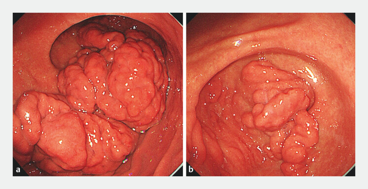 Fig. 1
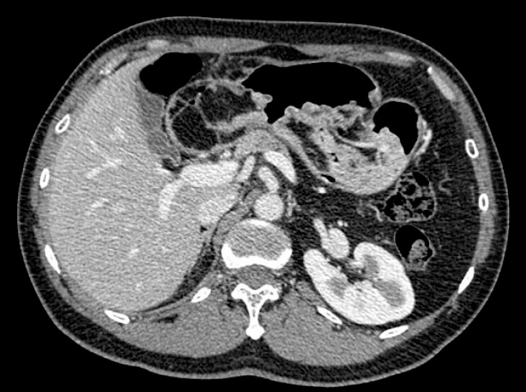 Fig. 2
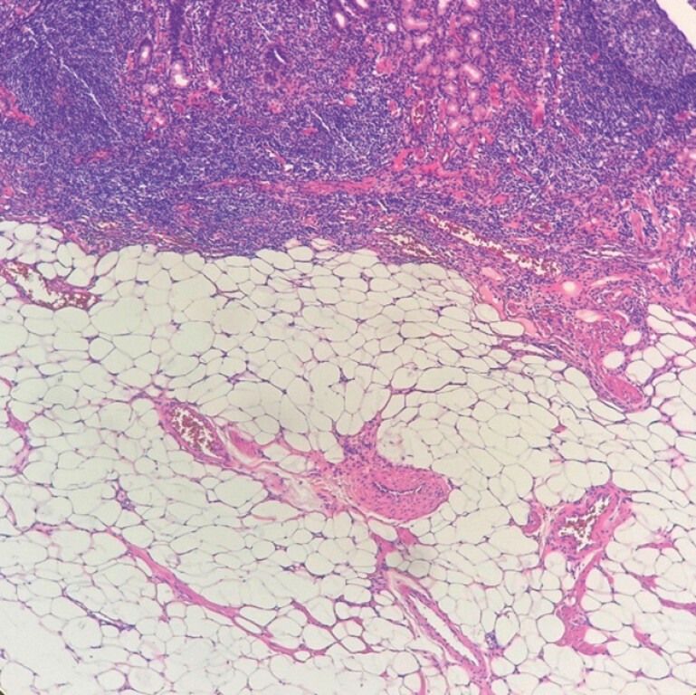 Fig. 3
- —The major technology application and demonstration project, Chengdu Science and Technology Bureau
Peer Reviews
No public reviews on file for this paper yet. If you reviewed it on a platform where reviews are public (OpenReview, ICLR, NeurIPS, ICML), you can paste yours below so the community can read it here.
Videos
No videos yet. Explain this paper in a talk, walkthrough, or lecture? Add one.
Taxonomy
TopicsGastrointestinal disorders and treatments · Gastrointestinal Tumor Research and Treatment · Gastric Cancer Management and Outcomes
Lipomas are benign tumors composed of mature adipocytes. Gastrointestinal (GI) lipomas are rare neoplasms, particularly those occurring in the stomach, which account for only 5% of all GI lipomas and 2%–3% of all benign gastric tumors. They are most commonly found in the gastric antrum 1 2 . Gastric lipomas are typically small and asymptomatic; however, as they enlarge, symptoms such as epigastric discomfort, gastric outlet obstruction, and dyspepsia may occur 3 .
We report the case of a 47-year-old man who was referred to our department following the detection of a large submucosal protrusion during an upper GI endoscopy at an external hospital. Esophagogastroduodenoscopy revealed a giant subepithelial lesion ( Fig. 1 ). Extensive, thickened, fold-like and nodular mucosal elevations resembling cerebriform changes were observed from the greater curvature of the lower gastric body to the antrum. A prominent protrusion extended into the duodenal bulb through the pylorus, with smooth overlying mucosa and a soft texture. Endoscopic ultrasonography demonstrated that the lesion was primarily located in the submucosa, with localized thickening up to 1.6 cm, exhibiting homogeneous hyperechogenicity. Elastography indicated a soft consistency, consistent with the characteristics of a lipoma. Contrast-enhanced computed tomography revealed nodular and mass-like lesions with fat density in the gastric body, antrum, and adjacent duodenal region, with the largest measuring approximately 7.2 × 5.8 cm, suggestive of a lipoma ( Fig. 2 ).
Endoscopic findings. a Giant cerebriform submucosal lipoma in the gastric body. b Giant cerebriform submucosal lipoma in the gastric antrum.
Contrast-enhanced computed tomography showed fat-dense nodules.
Endoscopic snare electrocautery resection was performed to remove two large tissue specimens for pathological examination, with no postoperative bleeding observed at the resection site ( Video 1 ).
A giant cerebriform submucosal lipoma extending from the gastric body to the duodenal bulb.Video 1
Histopathological examination of the biopsy specimens confirmed the presence of mature adipose tissue in the lamina propria and submucosa, supporting the diagnosis of a lipoma ( Fig. 3 ).
Histological analysis revealed mature fat tissue in the lamina propria and submucosa, diagnostic of lipoma (hematoxylin and eosin ×100).
To our knowledge, this is the first reported case of a giant gastric lipoma exhibiting cerebriform morphology and extending from the gastric body to the duodenal bulb. Although the lesion was extensive, the intact mucosa and absence of malignant features supported a benign diagnosis. Conservative management with regular follow-up may be considered; however, endoscopic resection should be performed if symptoms such as obstruction, bleeding, or ulceration develop 4 .
Endoscopy_UCTN_Code_CCL_1AB_2AD_3AB
The reference list from the paper itself. Each links out to its DOI / PubMed record.
- 1Cappell MS Stevens CE Amin M Systematic review of giant gastric lipomas reported since 1980 and report of two new cases in a review of 117110 esophagogastroduodenoscopies World J Gastroenterol 201723561928852321 10.3748/wjg.v 23.i 30.5619 PMC 5558125 · doi ↗ · pubmed ↗
- 2Sullivan IW Hota P Dass C Gastric lipomas: a case series and review of a rare tumor BJR Case Rep 201952.0180109 E 710.1259/bjrcr.20180109 PMC 672618331501708 · doi ↗ · pubmed ↗
- 3Krasniqi AS Hoxha FT Bicaj BX Symptomatic subserosal gastric lipoma successfully treated with enucleation World J Gastroenterol 200814593018855998 10.3748/wjg.14.5930 PMC 2751909 · doi ↗ · pubmed ↗
- 4Deprez PH Moons LMGOʼToole D Endoscopic management of subepithelial lesions including neuroendocrine neoplasms: European Society of Gastrointestinal Endoscopy (ESGE) Guideline Endoscopy 20225441242910.1055/a-1751-574235180797 · doi ↗ · pubmed ↗