Novel approach in the resection of oral maxillary tumours
Pablo Cea-Arestín, Arturo Bilbao-Alonso, Abel García-García, Carolina Menéndez-Lago

TL;DR
This paper introduces a new surgical technique using a temporal muscle flap to improve tumor removal and reconstruction in the oral maxillary area.
Contribution
The novel approach uses a temporal muscle flap for better tissue reconstruction and reduced surgical steps in oral maxillary tumor cases.
Findings
11 successful cases were treated using the temporal flap technique.
The technique offers shorter execution time and fewer surgeries, reducing patient morbidity and costs.
Abstract
Oral cavity tumours represent a global oral health issue due to their prevalence and incidence, in this paper, we will explain the procedure for the removal of various tumours and the restoration of the patient's anatomy, function and aesthetics through a novel approach that rely on a temporal muscle flap for the reconstruction of soft tissues in the surgical area. This study is based on an analysis of temporal flap technique data, collected at the public health hospital of Santiago de Compostela (CHUS), from 2015 to 2024. The results are shown in Table 1, being the most relevant data the fact of having a series of 11 successful cases treated with temporal flap. The surgical technique involves tumor resection, preferably through an intraoral approach. This novel technique must be taken into account when solving this type of oncological cases with associated maxillectomy due to it’s…
Genes, proteins, chemicals, diseases, species, mutations and cell lines named across the full text — each resolved to its canonical identifier and authoritative record.
Click any figure to enlarge with its caption.
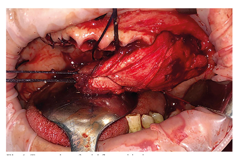 Figure 1
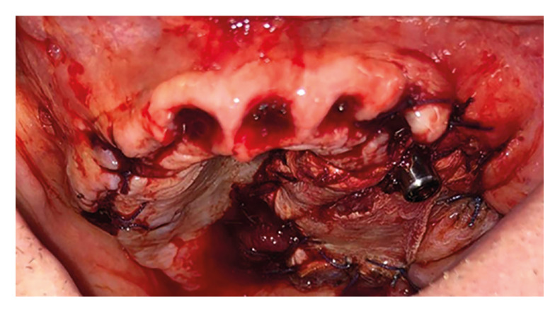 Figure 2
Peer Reviews
No public reviews on file for this paper yet. If you reviewed it on a platform where reviews are public (OpenReview, ICLR, NeurIPS, ICML), you can paste yours below so the community can read it here.
Videos
No videos yet. Explain this paper in a talk, walkthrough, or lecture? Add one.
Taxonomy
TopicsOral and Maxillofacial Pathology · Head and Neck Surgical Oncology · Oral and gingival health research
Introduction
Oral cavity tumours represent a global oral health issue due to their prevalence and incidence (1), with data showing that in 2022 the number of new cases detected reached 389,485 bringing a mortality rate of nearly 50% (2).
In this paper, we will explain the procedure for the removal of various tumours and the restoration of the patient's anatomy, function and aesthetics through a novel approach supported by the success of several performed cases.
After tumour removal, we will rely on a temporal muscle flap for the reconstruction of soft tissues in the surgical area along with the use of conventional dental, zygomatic and pterygoid implants, all of which have shown high success rates in such situations (3).
In particular, zygomatic implants have been proved especially useful, as one of their main indications is their use after maxillectomies due to oncologic causes (4-7).
Material and Methods
This study is based on an analysis of temporal flap technique data, collected at the public health hospital of Santiago de Compostela (CHUS), from 2015 to 2024.
The data coding, processing and statistical analysis were conducted using Excel as the primary computational and mathematical tool.
All ethical, healthcare, legal, data management and administrative requirements were strictly adhered to and in accordance with current regulations.
Results
The results are shown in Table 1, being the most relevant data the fact of having a serie of 11 successful cases.
A total of 11 patients were treated using this technique, temporal flap taken in 11 cases (100%), immediate implants were placed in 4 cases (36,36%) and the number of zygomatic implants positioned per case were 1 in 8 cases (72,73%) and 2 in 3 cases (27,27%).
The surgical technique involves tumor resection, preferably through an intraoral approach, in cases where this is not possible, a Weber-Ferguson approach will be used. After marking the resection area, the tooth in the premaxilla contiguous to the resective margin is extracted to allow the implant to be placed in the most medial location, based on findings from the finite element load distribution study (7-8). The osteotomy, usually up to the nasal septum to ensure the medial margin, is completed using piezoelectric instruments.
To harvest the temporal muscle graft, we approach the temporal area itself following the hairline to avoid potential areas of alopecia and the dissection of the temporal fascia is then performed lifting it carefully to include the facial nerve to prevent damage. Once reaching the bony plane at the temporal fossa, zygomatic arch, and frontomalar region, the temporal myofascial flap is detached and passed under the zygomatic arch, if the passage is too narrow or the flap is too large, osteotomy followed by osteosynthesis may be required (Fig. 1). This procedure serves as an alternative to harvesting the flap from the arm (9), thus resulting in lower morbidity.
Figure 1. Temporal myofascial flap positioning.
Once the flap is positioned in the oral cavity, the implants are placed, aiming to position at least one medial implant, one zygomatic implant, and, if oncologically feasible, a pterygoid implant (4,7), with the temporal flap ultimately positioned over the implants (Fig. 2).
Figure 2. Zygomatic implant surrounded by temporal flap.
Discussion
The indications for this type of surgery include: patients without prior reconstruction following a maxillectomy, patients with soft tissue reconstruction without bone reconstruction or implants, and patients undergoing simultaneous resection, reconstruction, and implant placement. The effectiveness and predictability of this procedure have been proven, with high implant survival rates (10).
The next step in this technique would be to make it a guided process with a fully digital workflow for maximum precision, this is feasible since there are currently digital guided methods with slight variability that still allow for prosthesis placement in the same procedure (11).
Conclusions
This novel technique must be taken into account when solving this type of oncological cases with associated maxillectomy due to it’s better results in terms of reconstruction of tissues and anatomy, shorter execution time and possibility of offering immediate function to the patient, leading to less number of surgeries with their consequent lower morbidity and social-health and economic savings.
The reference list from the paper itself. Each links out to its DOI / PubMed record.
- 1Kijowska J Grzegorczyk J Gliwa KJędras A Sitarz M Epidemiology, Diagnostics, and Therapy of Oral Cancer-Update Review Cancers (Basel)20241631563933512810.3390/cancers 16183156 PMC 11430737 · doi ↗ · pubmed ↗
- 2Bray F Laversanne M Sung H Ferlay J Siegel RL Soerjomataram I Global cancer statistics 2022: GLOBOCAN estimates of incidence and mortality worldwide for 36 cancers in 185 countries CA Cancer J Clin 202474229633857275110.3322/caac.21834 · doi ↗ · pubmed ↗
- 3Cea-Arestín P Bilbao-Alonso A Hernández-De Oliveira M Retrospective study of a serie of pterygoid implants Med Oral Patol Oral Cir Bucal 202429 e 650543890764510.4317/medoral.26633 PMC 11365061 · doi ↗ · pubmed ↗
- 4Weischer T Schettler D Mohr C Titanium implants in the zygoma as retaining elements after hemimaxillectomy Int J Oral Maxillofac Implants 199712211149109271 · pubmed ↗
- 5Tamura H Sasaki K Watahiki R Primary insertion of implants in the zygomatic bone following subtotal maxillectomy Bull Tokyo Dent Coll 2000412141121238010.2209/tdcpublication.41.21 · doi ↗ · pubmed ↗
- 6Grecchi F Bianchi AE Siervo S Grecchi E Lauritano D Carinci FA new surgical and technical approach in zygomatic implantology Oral Implantol (Rome)2017101972082987604510.11138/orl/2017.10.2.197PMC 5965068 · doi ↗ · pubmed ↗
- 7Ujigawa K Kato Y Kizu Y Tonogi M Yamane GY Three-dimensional finite elemental analysis of zygomatic implants in craniofacial structures Int J Oral Maxillofac Surg 200736620251751749710.1016/j.ijom.2007.03.007 · doi ↗ · pubmed ↗
- 8Ishak MI Abdul Kadir MR Sulaiman E Abu Kasim NH Finite element analysis of different surgical approaches in various occlusal loading locations for zygomatic implant placement for the treatment of atrophic maxillae Int J Oral Maxillofac Surg 2012411077892257517910.1016/j.ijom.2012.04.010 · doi ↗ · pubmed ↗