A rare clinical image of a 16-year-old adolescent with juvenile spondyloarthritis
Neha Ashok Brahmane, Sharath Hullumani

Abstract
Genes, proteins, chemicals, diseases, species, mutations and cell lines named across the full text — each resolved to its canonical identifier and authoritative record.
Click any figure to enlarge with its caption.
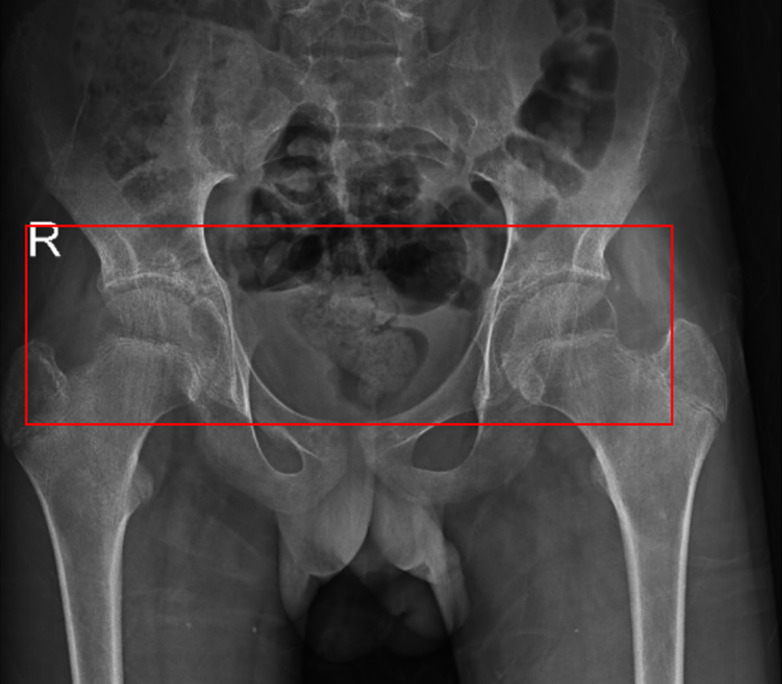 Figure 1
Peer Reviews
No public reviews on file for this paper yet. If you reviewed it on a platform where reviews are public (OpenReview, ICLR, NeurIPS, ICML), you can paste yours below so the community can read it here.
Videos
No videos yet. Explain this paper in a talk, walkthrough, or lecture? Add one.
Taxonomy
TopicsHip disorders and treatments · Infectious Diseases and Tuberculosis · Spondyloarthritis Studies and Treatments
Image in medicine
Juvenile spondyloarthritis is a chronic inflammatory condition that starts during childhood (before 16 years of age). Majorly characterizes arthritis, HLA-B27 positively, sacroiliitis, and inflammatory bowel disease. The prevalence estimates range from 1-4, which is 1% of the population. A 16-year-old boy visited a tertiary care hospital with complaints of severe pain in the hip joint while at rest as well as during movement. According to the primary caregiver, the child had a history of a fall from a bicycle on the right side of the body 5 years ago. The pain was progressive and later affected the activities of daily living. The magnetic resonance imaging (MRI) scan indicated bilateral hip arthritis with bilateral sacroiliitis. Subsequently, the patient underwent conservative treatment including intravenous antibiotics, antacids, antiemetics, analgesics, and various supportive treatments. For further rehabilitation, a structured physiotherapy program focused on pain management through early mobilization, reach-out exercises, and gait training. The case highlights the importance of combining medical treatment with structured physiotherapy rehabilitation to inhibit pain, optimize activities of daily living (ADL´s), enhance recovery, and improve patient´s well-being.
magnetic resonance imaging scan indicated bilateral hip arthritis with bilateral sacroiliitis, T2/PDFS hyperintensity was noted in the bilateral sacroiliac joint, erosion of bilateral acetabular surface, minimal bilateral hip joint effusion with marginal erosion along the articular surface, T2/PDFS hyperintensity was noted in the neck of the femur and greater trochanter of bilateral femur S/O edema