Isolated complete oculomotor nerve palsy as a presentation of adult medulloblastoma
Rafael Tuzino Leite Neves Maffei, Bruna Gutierres Gambirasio, Murillo Silva Catito, Sebastião Boanerges de Araujo Neto, Adrialdo José Santos

Abstract
Genes, proteins, chemicals, diseases, species, mutations and cell lines named across the full text — each resolved to its canonical identifier and authoritative record.
Click any figure to enlarge with its caption.
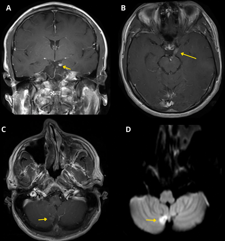 Figure 1
Peer Reviews
No public reviews on file for this paper yet. If you reviewed it on a platform where reviews are public (OpenReview, ICLR, NeurIPS, ICML), you can paste yours below so the community can read it here.
Videos
No videos yet. Explain this paper in a talk, walkthrough, or lecture? Add one.
Taxonomy
TopicsOphthalmology and Eye Disorders · Glioma Diagnosis and Treatment · Pituitary Gland Disorders and Treatments
A 37-year-old man presented with isolated complete oculomotor nerve palsy, without other neurological signs. Magnetic resonance imaging (MRI) showed thickening and enhancement of the left third cranial nerve, along with a small nodular lesion in the right cerebellar hemisphere with diffusion restriction ( Figure 1 ). Repeated cerebrospinal fluid analysis eventually confirmed the presence of neoplastic cells. Resection of the cerebellar lesion revealed desmoplastic/nodular-type medulloblastoma (MB), sonic hedgehog (SHH)-activated, and TP53 wildtype. The patient was treated with neuroaxis radiotherapy and adjuvant chemotherapy. This case demonstrates a rare presentation of adult MB, highlighting the importance of considering leptomeningeal dissemination in the differential diagnosis of isolated cranial neuropathies. 1 2 3 4
Magnetic resonance scan of the brain of the patient with medulloblastoma and cerebrospinal fluid dissemination. ( A ) Coronal and ( B ) Axial T1-weighted SE after gadolinium (Gd) shows thickening and contrast enhancement of the left oculomotor nerve (arrow). ( C ) Axial T1-weighted SE after Gd reveals a nodular lesion in the lower medial region of the right hemisphere of the cerebellum (arrow). ( D ) Axial DWI B1000 sequence shows restricted diffusion in the nodular lesion (arrow).
The reference list from the paper itself. Each links out to its DOI / PubMed record.
- 1Majd N K Mastall M Lin H Dibaj S S Hess K R Yuan Y Clinical characterization of adult medulloblastoma and the effect of first-line therapies on outcome; The MD Anderson Cancer Center experience Neurooncol Adv 2021301 vdab 07910.1093/noajnl/vdab 07934377987 PMC 8350154 · doi ↗ · pubmed ↗
- 2Cocito C Martin B Giantini-Larsen A M Valcarce-Aspegren M Souweidane M M Szalontay L Leptomeningeal dissemination in pediatric brain tumors Neoplasia 20233910089810.1016/j.neo.2023.10089837011459 PMC 10124141 · doi ↗ · pubmed ↗
- 3Russo C Scala M R Spennato P Nastro A Errico M E De Martino L Cinalli G Primary leptomeningeal medulloblastoma: a case-based review Childs Nerv Syst 2022380352753610.1007/s 00381-021-05435-x 35059784 · doi ↗ · pubmed ↗
- 4Zhao F Li C Zhou Q Qu P Wang B Wang X Distinctive localization and MRI features correlate of molecular subgroups in adult medulloblastoma J Neurooncol 20171350235336010.1007/s 11060-017-2581-y 28808827 · doi ↗ · pubmed ↗