Worsening Hiccups, Dyspnea, and Angina in a 67-Year-Old Woman: A Challenging Case
Stefanos Votsis, Jaime Caballero, Cezar Iliescu, Konstantinos Marmagkiolis

TL;DR
A rare heart condition called left atrial appendage aneurysm is presented in a 67-year-old woman with worsening symptoms, emphasizing the need for early surgical treatment.
Contribution
Highlights the importance of early surgical intervention for left atrial appendage aneurysm to prevent serious complications.
Findings
Left atrial appendage aneurysm can present with hiccups, dyspnea, and angina.
Early surgical intervention is recommended to prevent thromboembolic complications and arrhythmias.
Abstract
In this publication, we present a left atrial appendage aneurysm (LAAA) case diagnosed with a cardiac CT scan of a 67-year-old woman with worsening hiccups, dyspnea, and chest pain. This is a rare cardiac condition with only 150 cases reported to date. LAAA can manifest as diastolic dysfunction, angina, hiccups, arrhythmias, dyspnea, and rare but potentially serious complications such as systemic embolism and rupture leading to death. Our findings highlight the importance of early surgical intervention, even in asymptomatic patients, to mitigate potential thromboembolic complications and address associated atrial arrhythmias.
Genes, proteins, chemicals, diseases, species, mutations and cell lines named across the full text — each resolved to its canonical identifier and authoritative record.
Click any figure to enlarge with its caption.
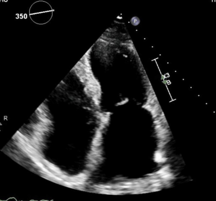 Figure 1
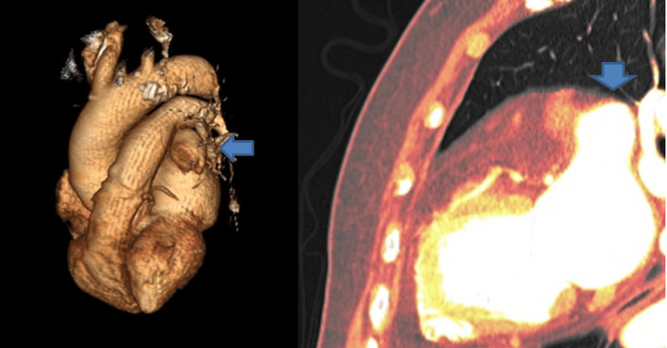 Figure 2
Peer Reviews
No public reviews on file for this paper yet. If you reviewed it on a platform where reviews are public (OpenReview, ICLR, NeurIPS, ICML), you can paste yours below so the community can read it here.
Videos
No videos yet. Explain this paper in a talk, walkthrough, or lecture? Add one.
Taxonomy
TopicsPathogenesis and Treatment of Hiccups · Urticaria and Related Conditions · Heparin-Induced Thrombocytopenia and Thrombosis
Introduction
Anatomic variations of various cardiac structures are not always apparent and might prove challenging to diagnose [1]. Initial suspicion usually depends on clinical symptoms, which are oftentimes non-specific. Various cardiac imaging entities may prove extremely useful in reaching diagnosis, as well as tailoring the appropriate therapeutic management of these patients.
One of the most uncommon conditions that has garnered attention from the medical community due to its low incidence and varied clinical manifestations is the left atrial appendage aneurysm (LAAA). The difficulty in identification is reflected in its incidental detection in imaging studies such as echocardiograms (both transthoracic and transoesophageal) and tomographies (both computer and magnetic resonance), while symptoms range from mild to severe, including heart failure and thromboembolic events. The complex etiology includes congenital and acquired factors, and its management focuses on preventing complications through surgical resection, accompanied by medical strategies such as controlling heart rhythm and anticoagulation [2].
Case presentation
A patient in her mid-60s with a medical history of transient ischemic attack (TIA) and palpitations presented with worsening hiccups, dyspnea, and angina. Physical examination and bloodwork results were unremarkable. A chest X-ray, transthoracic echocardiogram (Figure 1), and a pharmacologic nuclear stress test were normal.
Transthoracic echocardiogram image
A two-week monitor showed only rare premature ventricular contractions (PVCs). Lacking a diagnosis explaining the patient's symptoms, a further decision was made to order a cardiac CT, whose result is shown in Figure 2.
Cardiac CT 3D reconstruction image; left atrial appendage aneurysm (blue arrow)
Discussion
The cardiac CT scan demonstrates a left atrial appendage aneurysm (LAAA) (Figure 2, blue arrow) whose dimensions are measured at 6x3x3 cm, with no apparent thrombus. First documented in 1962 [3,4], approximately 150 cases have been reported in the literature to date [5]. LAAA can manifest across all age groups, with a mean age of diagnosis at 30 years. Approximately 90% of cases are congenital, associated with congenital dysplasia of the atrial pectinate muscles. The common histopathological finding is fibrosis of the endocardium or myocardium.
LAAA can lead to various clinical manifestations, including diastolic dysfunction resulting from compression of the left ventricle, angina induced by compression of the left anterior descending (LAD) coronary artery, hiccups due to irritation of the left phrenic nerve [6], arrhythmias (most commonly atrial fibrillation, atrial flutter, and supraventricular tachycardia), dyspnea accompanied by a dry, unproductive cough stemming from irritation of the respiratory tract, systemic embolism, and, in rare instances, death attributable to rupture [7].
Imaging modalities that assist the physician in the diagnosis of LAAA are chest X-ray and cardiac ultrasound (both transthoracic and transesophageal), although tomographic studies, as well as magnetic resonance imaging, play a crucial role in diagnosing and characterizing aneurysmal lesions of the cardiac cavities. Interestingly, more patients are reported to undergo cardiac CT imaging than transesophageal cardiac ultrasound (TOE) (46% vs 40%) [7], and this is also the reason for our prioritizing cardiac CT over TOE in our diagnostic algorithm.
Surgical intervention is the main treatment modality for LAAA, whether symptomatic or not, in order to prevent potential complications, and is performed in more than 75% of the patients [7]. Various effective techniques, including median sternotomy, left thoracotomy, mini-thoracotomy, and minimally invasive endoscopic approaches, have been reported for aneurysmatectomy. Our patient underwent surgical resection of the LAAA, with a favorable result.
Conclusions
Rare clinical entities are always a part of the clinician’s diagnostic algorithm. Modern diagnostic modalities are invaluable and can prove very helpful in the resolution of the most challenging of cases.
Left atrial appendage aneurysm is one such rare clinical entity that can lead to arrhythmias and thromboembolic events. Surgical resection appears to be the primary treatment option in the current literature, and most patients show improvement or are asymptomatic after treatment. Additionally, alternative approaches, such as transcatheter closure of LAAA, ablation, and medical treatments, have been reported as viable alternatives to surgical intervention.
The reference list from the paper itself. Each links out to its DOI / PubMed record.
- 1Anatomic variants mimicking pathology on echocardiography: differential diagnosis J Cardiovasc Ultrasound Kim MJ Jung HO 1031122120132419891510.4250/jcu.2013.21.3.103PMC 3816159 · doi ↗ · pubmed ↗
- 2Left atrial appendage aneurysm: a case report and literature review Cureus Ayala Torres JD Sepulveda Gallego JA Gonzalez Gonzalez M 016202410.7759/cureus.56280 PMC 1101800938623095 · doi ↗ · pubmed ↗
- 3Congenital atriomegaly Circulation Parmley LF Jr 5535582519621448398710.1161/01.cir.25.3.553 · doi ↗ · pubmed ↗
- 4Left atrial appendage aneurysm: a systematic review of 82 cases Echocardiography Aryal MR Hakim FA Ghimire S 131213183120142497637610.1111/echo.12667 · doi ↗ · pubmed ↗
- 5Left atrial appendage aneurysm: a case report World J Clin Cases Belov DV Moskalev VI Garbuzenko DV Arefyev NO 44434449820203308340310.12998/wjcc.v 8.i 19.4443 PMC 7559682 · doi ↗ · pubmed ↗
- 6Huge left atrial appendage aneurysm revealed by chronic hiccups J Saudi Heart Assoc Asfalou I Boumaaz M Raissouni M Sabry M Benyass A Zbir EM 2932962920172898317310.1016/j.jsha.2017.03.009PMC 5623021 · doi ↗ · pubmed ↗
- 7Left atrial appendage aneurysm: a descriptive systematic review of 177 cases BMC Cardiovasc Disord Daralammouri Y Odeh A Abuzahra S Azamtta M Shawahna R 6332420243952894610.1186/s 12872-024-04323-x PMC 11552148 · doi ↗ · pubmed ↗