Correction: Ginsenoside compound K attenuates Ox-LDL-mediated macrophage inflammation and foam cell formation via autophagy induction and modulating NF-κB, p38, and JNK MAPK signaling
Shan Lu, Yun Luo, GuiBo Sun, XiaoBo Sun

Abstract
Genes, proteins, chemicals, diseases, species, mutations and cell lines named across the full text — each resolved to its canonical identifier and authoritative record.
Click any figure to enlarge with its caption.
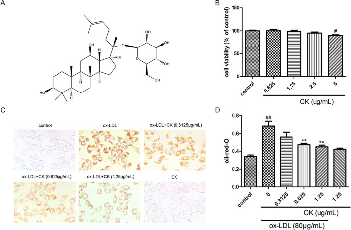 Figure 1
Peer Reviews
No public reviews on file for this paper yet. If you reviewed it on a platform where reviews are public (OpenReview, ICLR, NeurIPS, ICML), you can paste yours below so the community can read it here.
Videos
No videos yet. Explain this paper in a talk, walkthrough, or lecture? Add one.
Taxonomy
TopicsGinseng Biological Effects and Applications · Cancer-related molecular mechanisms research · Lipid metabolism and disorders
In the published article, there was an error in the legend for Figure 1 as published. In Figure 1D, it was an error to state that oil red O positive area was measured by Image J software. The corrected legend appears below.
“CK inhibited ox-LDL-induced RAW264.7 cells lipid accumulation. RAW264.7 cells were treated with CK at various concentrations for 12 h with or without 80 μg/mL ox-LDL for additional 24 h. (A) The chemical formula for CK. (B) Cell viability was assayed by the MTT assay. (C) Representative images of Oil Red O staining. (D) OD value results of oil red O. All data are shown as mean ± SD from three independent experiments with each performed in triplicate. ^#^ P < 0.05, ^ ## ^ P < 0.01 vs. control group; **P < 0.01 vs. ox-LDL-treated group. CK, compound K; ox-LDL, oxidized low-density lipoprotein; MTT, (4, 5-dimethylthiazol-2yl-)-2,5-diphenyl tetrazolium bromide.”
In the published article, there was an error in Figure 1 as published. In Figure 1C, the representative picture of the CK group was updated with the correct one. The corrected Figure 1 and its caption appear above.
In the published article, there was an error in the legend for Figure 6 as published. In Figure 6D, it was an error to state that oil red O positive area was measured by Image J software. The corrected legend appears below.
“CK mediated-autophagy and anti-inflammation were abolished by NF-κB, P38, and JNK MAPK activation. RAW264.7 cells were treated with CK (1.25 μg/mL) for 12 h with or without the NF-κB inhibitor, PDTC (10 μM) or the MAPK activator, anisomycin (0.1 μM) or the autophagy inhibitor 3-MA (5 mM). Then cells were stimulated with 80 μg/mL ox-LDL for 24 h. (A) The protein expression levels of LC3, Beclin-1, P62, IL-1β, TNF-α, and β-actin were examined by western blot assay. (B) Statistical results of LC3II/LC3I, Beclin-1, P62, IL-1β, and TNF-α expression levels. (C) Representative images of Oil Red O staining. (D) OD value results of oil red O. (E) Representative Western blot analysis of phosphorylated and total p38, and JNK was performed. (F) The expression levels of LC3, Beclin-1, P62, IL-1β, TNF-α, and β-actin were detected by Western blot analysis. (G) Densitometric analysis was used to quantify the levels of p-p38, p-JNK. (H) Statistical results of LC3II/LC3I, Beclin-1, P62, IL-1β, and TNF-α expression levels. All data are shown as mean ± SD from three independent experiments with each performed in triplicate. ^ # ^ P < 0.05, ^ ## ^ P < 0.01, ^ ### ^ P < 0.001 vs. control group; *P < 0.05, **P < 0.01, ***P < 0.001 vs. ox-LDL-treated group; ^ & ^ P < 0.05, ^ && ^ P < 0.01 vs. ox-LDL and CK treatment group. CK, compound K; PDTC, pyrrolidinedithiocarbamate ammonium; 3-MA, 3-Methyladenine; AM, anisomycin.”
The original version of this article has been updated.