N‑Doped Carbon Dot-Based Nanoconjugates with Simultaneous Generation of Nitric Oxide and Singlet Oxygen for Phototherapeutic Applications
Francesca Laneri, Cristina Parisi, Vittoria Andrigo, Juliana Guerra Pinto, Luciana Cortez Marcolino, Juliana Ferreira-Strixino, Marta Maria Natile, Salvatore Sortino

TL;DR
This paper introduces a new nanoconjugate that generates both nitric oxide and singlet oxygen under green light for enhanced cancer phototherapy.
Contribution
The novel nanoconjugate combines N-doped carbon dots with a NO photodonor, enabling simultaneous generation of NO and singlet oxygen under green light.
Findings
The nanoconjugate allows intramolecular photoinduced electron transfer for NO release with improved excitation wavelength.
Green light excitation generates singlet oxygen via collisional energy transfer with molecular oxygen.
The nanoconjugate localizes in the cytoplasm of brain cancer cells and increases cell mortality under green light.
Abstract
Nitric oxide (NO) and singlet oxygen (1O2) represent two of the most intriguing agents for unconventional phototherapeutic applications in cancer. In this contribution, N-doped carbon dots (NCDs) with strong absorption in the biocompatible green region have been synthesized and covalently decorated with an otherwise blue-light-activatable NO photodonor (NOPD), leading to a nanoconjugate ca. 3.5 nm in diameter. The NCD core of the nanoconstruct acts as a green light antenna, permitting the release of NO from the NOPD by an intramolecular photoinduced electron transfer, with an improvement of more than 100 nm in the excitation wavelength. Simultaneously, green light excitation generates 1O2 by collisional energy transfer with molecular oxygen. Due to its emissive properties, the nanoconjugate can be visualized in 9L/LacZ brain cancer cells, where it localizes mainly in the cytoplasm.…
Genes, proteins, chemicals, diseases, species, mutations and cell lines named across the full text — each resolved to its canonical identifier and authoritative record.
Click any figure to enlarge with its caption.
 1
1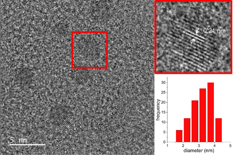 1
1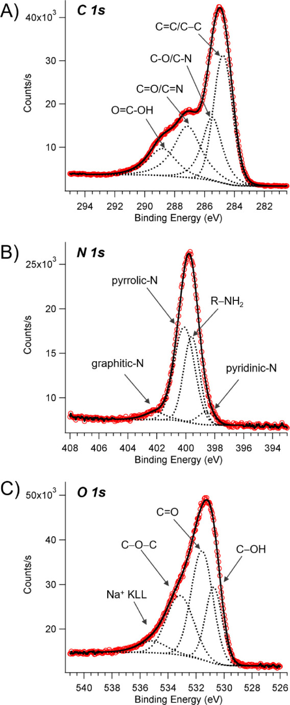 2
2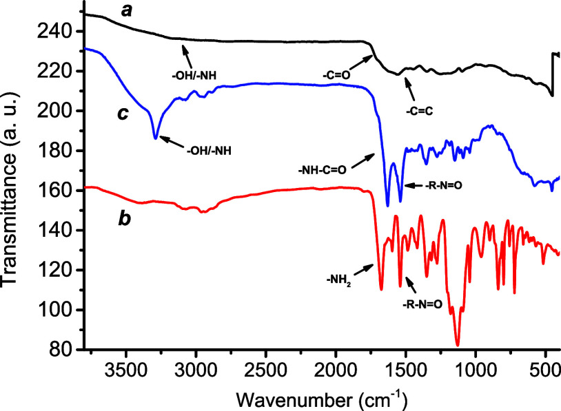 3
3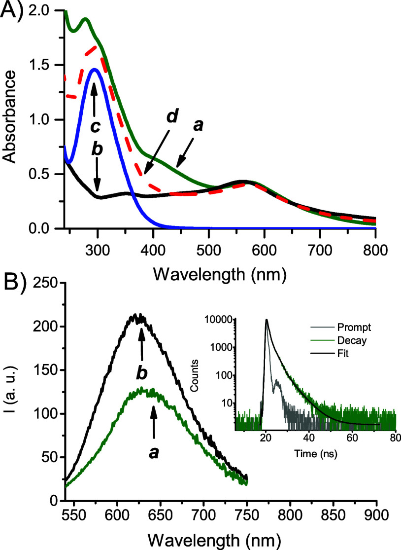 4
4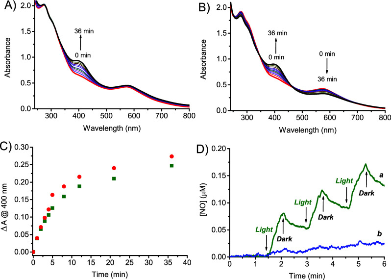 5
5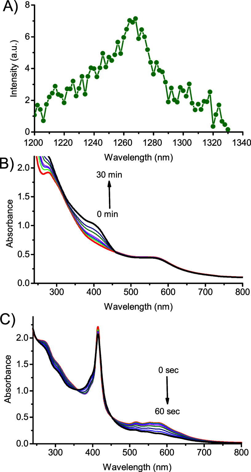 6
6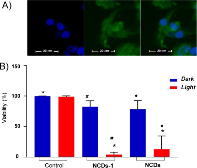 7
7- —Associazione Italiana per la Ricerca sul Cancro10.13039/501100005010
- —Ministero dell'Universit? e della Ricerca10.13039/501100021856
Peer Reviews
No public reviews on file for this paper yet. If you reviewed it on a platform where reviews are public (OpenReview, ICLR, NeurIPS, ICML), you can paste yours below so the community can read it here.
Videos
No videos yet. Explain this paper in a talk, walkthrough, or lecture? Add one.
Taxonomy
TopicsCarbon and Quantum Dots Applications · Graphene and Nanomaterials Applications · Advanced Nanomaterials in Catalysis
Introduction
1
Nitric oxide (NO) and singlet oxygen (^1^O_2_) represent two of the most intriguing unconventional agents for phototherapeutic applications. ?−? ? They combine several common advantages over conventional drug molecules, such as the absence of multidrug resistance, reactivity with all biological components, and, due to their short lifetime, confinement of their region of action to below 200 μm, with reduced systemic effects.
NO plays many physiological and pathophysiological roles, ?,? and its use as a therapeutic agent in several diseases including cancer has been extensively demonstrated. ?−? ? ? ? ? ? ? However, NO’s effects in cancer strictly depend on its concentration and generation site.? This makes the light-activated NO precursors, namely NO photodonors (NOPDs), very appealing. ?−? ? ? ? ? ? ? ? In NOPDs, the excitation light breaks a covalent bond, uncaging the NO that was initially integrated within their molecular skeleton.
^1^O_2_ plays a dominant role in photodynamic therapy (PDT) ?,? and, in contrast to NO, is usually generated in a catalytic fashion by suitable photosensitizers (PS) through collisional energy transfer with nearby molecular oxygen. ?−? ?
PSs and NOPDs present the great advantage of not being active in the dark but generating a burst of cytotoxicity exclusively under light inputs in the region of space confined to the irradiated area with superb spatiotemporal control. In this frame, creating single nanoplatforms that combine bimodal phototherapeutic performance and exploit the additive/synergistic effects of simultaneously generated NO and ^1^O_2_ has proven to be innovative in nanomedicine. ?−? ? ? Our pioneering studies in this regard ?,? have inspired the achievement of a number of molecular hybrids and supramolecular nanoconstructs devoted to this goal. ?,? Due to their covalent linking, the former ensure that both cytotoxic species are generated in the very same region of space but, on the other hand, present the limitation of a small reservoir of NO compared to the catalytically generated ^1^O_2_ due to the equimolar NOPD:PS molar ratio. The latter offer the advantage of facile tuning of the NOPD:PS molar ratio, but in contrast, these components can diffuse apart due to potential disassembling in a biological environment. On these bases, achieving robust nanostructures covalently integrating multiple NOPD and PS in the same scaffold is challenging since they can overcome the above limitations.
Carbon dots (CDs) are usually spherical carbon nanoparticles exhibiting low toxicity, excellent biocompatibility, and high cell permeability.? They consist of a core rich mainly of sp^2^ hybrid carbons and a shell that can bear different organic functional groups, including amines, hydroxyl, and carboxylic, depending on the precursors used for the synthesis. ?−? ? Besides making CDs well-dispersible in water, these moieties permit surface engineering with additional functional molecular units through simple synthetic protocols. ?−? ? CDs show excitation wavelength-dependent emission, which is helpful for their tracking in a bioenvironment, ?,? and can act as both electron/energy donors and acceptors in intra- and interphotoinduced processes with suitable counterparts. ?−? ? This wealth of properties makes these materials intriguing nanoplatforms for various applications in nanomedicine and photonanomedicine. ?−? ? ? In this regard, we have recently reported on N-doped CDs (NCDs) decorated with an NOPD activatable by blue light, demonstrating that blue light excitation amplifies the NO release from the NOPD through an intramolecular photoinduced electron transfer from the NCDs core to the peripheral NOPD.?
One of the most interesting aspects of CDs relates to their recently demonstrated capability to generate ^1^O_2_ as alternative to the typical PS based on either porphyrinoids or BODIPY derivatives. ?−? ?
NCDs have been revealed to be more efficient in this regard, with the photosensitization properties strictly dependent on the N doping.?
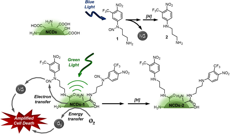
The above scenarios inspired us to achieve an NCDs-based nanoconstruct that can simultaneously generate NO and ^1^O_2_ with highly biocompatible green light. To this end, we have devised a novel nanoconjugate NCDs-1 (Scheme). It covalently integrates the NOPD 1, developed in our group ?,? and otherwise activatable by blue light, into NCDs exhibiting significant absorption in the green region. We show that the NCDs core of the nanoconstruct acts as the sole green light-harvesting antenna and triggers NO generation, probably by a photoinduced electron transfer with NOPD 1 in the shell, and ^1^O_2_ by a bimolecular energy transfer with molecular oxygen. This results in the amplified mortality of cancer cells due to the simultaneous photodynamic action of these two cytotoxic species.
Sketch of the Naked NCDs, the NOPD 1 and Its Stable Photoproduct 2 Formed after NO Release under Blue Light, and the Nanoconjugate NCDs-1 with its Working Principle.
Experimental Section
2
Materials and Methods
2.1
All chemicals were purchased from Sigma-Aldrich and used as received. All solvents used (Sigma-Aldrich) were of spectrophotometric grade. Deionized ultrafiltered water was used throughout this study.
Synthesis
2.1.1
NOPD 1 was synthesized as previously described. ?,?
NCDs were synthesized via an already reported solvothermal method using citric acid and urea, with dimethylformamide (DMF) as the solvent,? with some modifications. Specifically, 1 g of citric acid was reacted with 2 g of urea at 160 °C for 6 h in 10 mL of DMF. After cooling at room temperature, the resulting dark red solution was treated with 20 mL of an aqueous NaOH solution (50 mg mL^– 1^) and stirred for 1 min. The obtained solution was dialyzed overnight and later freeze-dried to give a dark purple product. 100 mg of this product was then solubilized in 20 mL of an aqueous HCl solution (5 wt %) and stirred for 10 min to remove surface metal cations. The solution was then centrifuged at 14,000 rpm for 10 min, and the solid was collected, solubilized in water, and centrifuged again twice at 14,000 rpm for 10 min, to eliminate residual salts and HCl. The final product was freeze-dried, yielding a dark powder of NCDs.
NCDs-1 were prepared as follows: To a 3 mL aqueous dispersion containing 15 mg of NCDs, 144 mg of 1-ethyl-3-(3-(dimethylamino)propyl)carbodiimide (EDC) and 86 mg of N-hydroxysuccinimide (NHS) were added. The mixture was sonicated for 15 min in an ice bath. Subsequently, 1 mL of an acetonitrile solution containing 45 mg of compound 1 was added, and the reaction mixture was stirred for 2 days in the dark at room temperature. Afterward, the acetonitrile was evaporated under vacuum, and the resulting product was dialyzed overnight and freeze-dried, yielding NCDs-1. Based on the molar absorptivity of compound 1 at 290 nm (9,600 M^–1^ cm^–1^), a functionalization degree of ca. 70% can be estimated.
Fluorescence, NO, and 1O2 Quantum Yields
2.1.2
Fluorescence quantum yields (Φ_f_) were determined at λ_exc_ = 530 nm using Rhodamine 6G in EtOH (Φ_f_ = 0.96) as the standard.?
NO photogeneration quantum yield (Φ_NO_) was determined at λ_exc_ = 532 nm, according to our reported procedure.?
^1^O_2_ quantum yields (Φ_Δ_) were determined at λ_exc_ = 532 nm in D_2_O (1% MeOD) using Rose Bengal as the standard (Φ_Δ_ = 0.76).?
Cell Experiments
2.2
Gliosarcomas of the 9L/LacZ lineage obtained from the Rio de Janeiro Cell Bank were maintained in Dulbecco’s Modified Eagle’s Medium (DMEM) (Gibco), supplemented with 10% fetal bovine serum (FBS) (LGC Biotechnology) and 1% penicillin/streptomycin solution (LGC Biotechnology), and kept in an incubator at 37 °C with 5% CO_2_.
Internalization
2.2.1
Initially, 10^6^ cells were adhered to glass coverslips disseminated in 24-well plates and left to incubate for 24 h in an incubator at 37 °C with 5% CO_2_. The culture medium was then removed, and NCDs-1 was added and incubated for 1 h, to allow the compound to be internalized, followed by washing with PBS and fixation with 4% paraformaldehyde for 15 min at room temperature. The slides were mounted with ProLong Diamond Antifade Mountant with DAPI (Thermo Fisher) and analyzed under a Zeiss LSM 700 confocal microscope. The groups were protected from light during the process.
Cell Viability
2.2.2
10^5^ cells were transferred in two 96-well plates, one plate was irradiated, and the other to remained in the dark. After cell adhesion, the medium was removed, and either NCDs or NCD-1 were added. For the groups without the compound, the same volume of PBS was added, and the plates were then irradiated in Biotable (Biotable PhotoBioS) for 48 min (530 nm, 83 mW/cm^2^). The Biotable Photobios GreenModel V1 (Photobios, Brazil) is a custom-designed photobiological irradiation platform equipped with 12 high-power LEDs (10 W each) emitting green light at a wavelength of 520 nm. The system provides a calibrated irradiance of 83.33 mW/cm^2^ over a uniform 11 × 16 cm active area, ensured by the fixed arrangement of the LEDs at 1 cm from the irradiation plane and a 120° emission angle per diode. After irradiation, the PBS and compounds were removed from the plates, and DMEM medium was added. The plates were incubated at 37 °C and 5% CO_2_ for 24 h. The experiment was performed in quadruplicate and protected from light during the process. After the respective treatments, viability tests were performed using the rypan blue assay. This method allows for the differentiation of live cells from dead cells by observing cell coloration. The procedure was performed 24 h after the application of treatment, with a 0.2% Trypan Blue solution (Sigma), and incubated for 5 min. After this time, the solution was removed, and PBS was added. The groups were analyzed using a Zeiss Axio Vert.A1 inverted microscope.
Statistical analysis
2.2.3
Statistical analyses were performed using GraphPad software, version 7.04 (GraphPad Software, Inc., La Jolla, CA, USA). Statistically significant comparisons between the experimental groups and the control group were presented, considering results with p < 0.001 as significant.
Instrumentations
2.3
HRTEM images were acquired with a previously described microscope.?
XPS measurements were performed with an ESCALAB QXi spectrometer from Thermo Fisher Scientific, using a monochromatic Al Kα source (1486.6 eV) operating at 200 W and a spot size of 650 μm × 200 μm.
The instrumentation for FTIR, UV–vis absorption and emission spectra, and time-resolved fluorescence has been previously described.?
Steady-state irradiation experiments were performed according to already reported setup? using a CW green laser (200 mW) at λ = 532 nm or, in the case of TPPS, with a LED at 420 nm (10 mW).
NO and ^1^O_2_ were detected by a direct method exploiting an ultrasensitive NO electrode and the typical NIR luminescence of ^1^O_2_, respectively, as previously reported. ?,?
Results and Discussion
3
HRTEM micrograph of NCDs reveals quite dispersed, spherical-like shaped NCDs with a mean diameter of 3.3 ± 0.1 nm (Figure). The NCDs are crystalline, as indicated by the well-visible lattice fringes. The interplanar spacing of 0.21 nm accords with that observed in graphene sheets.?
Representative HRTEM images and size distribution of NCDs.
The XPS analysis of NCDs reveals the presence of C (60.4 at%), N (24 at%), O (11.9 at%), and traces of Na (3.8 at%). Careful analysis of the C 1s, N 1s, and O 1s high-resolution spectra provided information on the chemical environment of different species (FigureA–C).
XPS analysis of NCDs,: high resolution spectra of (A) C 1s, (B) N 1s, and (C) O 1s. Red circles for raw data; black continuum line and dashed lines for the fitting curves.
The deconvolution of the C 1s core level reveals the presence of four different carbon species at 284.8 eV (CC, C–C), 285.5 eV, 287.1 eV (CO, CN), and 288.8 eV (OC–OH), respectively. ?,? The N 1s spectrum can be deconvoluted into four peaks with maxima at 398.6 eV, 399.6 eV, 400.1 eV, and 402.1 eV, consistent with pyridinic-N, aminic R-NH_2_, pyrrolic-N, and graphitic-N, respectively.? The O 1s region consists of three contributions at 530.7 eV, 531.6 eV, and 533.1 eV, ascribable to C–OH, CO, and O–C–O. The small peak at 535.1 eV is due to KLL of Na^+^.? The atomic percentages of different species are shown in Table S1.
FTIR analysis of NCDs (Figure) showed the presence of a broad band ranging from ca. 3700 cm^–1^ to 2900 cm^–1^, attributed to the typical stretching frequency of −OH, −NH, and −COOH groups localized on the surface scaffolds, responsible for the hydrophilic nature of the nanostructures,? which resulted in them being well dispersible in water and suitable for further functionalization. The band at 1555 cm^–1^ was related to CC stretching, and the distinctive band at 1700 cm^–1^ was related to the CO stretching of the carboxylic groups. FTIR analysis also proved the successful grafting of compound 1 at the NCDs shell (Figure). The peak related to the CO stretching shifted to 1632 cm^–1^ in NCDs-1, in good agreement with the formation of an amide bond. In contrast, the stretching vibration of −R–NO at 1540 cm^–1^ in the case of 1? was unaltered in the nanoconjugate. HRTEM analysis of NCDs-1 revealed no significant changes in size and morphology with respect to NCDs (Figure S1). NCDs-1 were well-dispersible in water medium, remaining stable for ca. one month with negligible evidence of aggregation (confirmed by the unchanged absorption spectrum, see below).
FTIR spectra of NCDs (a), 1 (b), and NCDs-1 (c).
Compound 1 exhibits an absorption maximum at 290 nm and a tail extending beyond 400 nm (Figure S2). Irradiation of compound 1 with blue light induces the homolytic rupture of the N–NO bond, decaging of NO, and formation of the stable photoproduct 2 after H transfer from the solvent to the anilinyl radical intermediate (see Scheme). ?,? The stable photoproduct 2 exhibits molar absorptivity similar to compound 1, but its absorption maximum is significantly shifted to longer wavelengths (ca. 100 nm) due to the push–pull character of the nitroaniline moiety (Figure S2). ?,?,?,?
FigureA shows the absorption spectra of NCDs-1 and, for the sake of comparison, those of NCDs, compound 1, and their mixture. NCDs-1 exhibits the fingerprints of both components with an intense UV band at ca. 300 nm, typical of compound 1, and the broad absorption of NCDs in the green spectral window at ca. 560 nm. Besides, a new absorption is present in the interval of 400–500 nm. These absorption features differ from those of the mixture of the two components (see spectrum d in FigureA) and account for a remarkable interaction between compound 1 and the NCDs scaffold of the nanoconjugate in the ground state. On the basis of the electron-accepting and electron-donating features of compound 1 and NCDs, respectively, the new absorption in the region of 400–500 nm is probably charge transfer in character. This is in agreement to what already found in our recent work for the same NOPD grafted onto UV-absorbing NCDs ? and is supported by the study of Guldi and coworkers in the case of nanoconjugates of CDs integrating electron-acceptor chromophoric components.?
(A) Absorption spectra of aqueous dispersions (1% MeOH) of NCDs-1 (70 μg mL–1) (a), NCDs (17 μg mL–1) (b), 1 (53 μg mL–1) (c), and the mixture of NCDs + 1 (d). (B) Fluorescence emission spectra of aqueous dispersions (1% MeOH) of NCDs-1 (70 μg mL–1) (a) and NCDs (17 μg mL–1) (b) at λexc = 530 nm, T = 25 °C. The inset shows the fluorescence decay and the related triexponential fitting of the NCDs.
As typically observed in different types of CDs,? NCDs show emission dependent on the excitation wavelength (Figure S3) and, in this case, it mainly falls in the green/red region. FigureB shows that the emission of NCDs (Φ_f_ = 0.03) is partially quenched in NCDs-1 (Φ_f_ = 0.018). These findings account for an interaction between the components in the excited state. Regarding the quenching observed, energy transfer between the NCDs core and the peripheral 1 is out of the question. The emission of the former (energy donor) falls, in fact, well beyond the absorption of the latter (energy acceptor) (absence of spectral overlap), making this process thermodynamically unfeasible. Instead, we believe that analogously to what was observed in our previous work,? a photoinduced electron transfer from NCDs to the strong electron acceptor 1 is more likely. Note that the emission dynamics of NCDs exhibit a triexponential decay, typical for CDs, ?,? with lifetimes (τ) and related amplitudes (α) being τ_1_ = 4.87 ns (α_1_ = 12.36%), τ_2_ = 1.8 ns (α_2_ = 39.22%), and τ_3_ = 0.29 ns (α_3_ = 48.41%) (inset FigureB) and attributable to diverse fluorophoric domains. ?,? These values did not significantly change in NCDs-1, suggesting that the quenching of the emission takes place on a time scale below our time resolution (ca. 200 ps). This hypothesis is in line to what is already found in CD nanoconjugates functionalized with strong electron acceptor chromophoric components, in which quenching by photoinduced electron transfer occurring on a ps time scale was observed by ultrafast spectroscopy.?
Irradiation of NCDs-1 with green light under anaerobic conditions shows that the absorption of the NCDs core beyond 560 nm remains almost unaltered, whereas a new absorption band is observed at ca. 400 nm (FigureA). The growth of this band is also observed under aerobic conditions, but in this case, bleaching of the NCDs core absorption is noted (FigureB). The kinetic profile at 400 nm is very similar in the absence or presence of oxygen (FigureC). Note that the new band at ca. 400 nm is the same as that observed for the unbound 1 under blue light excitation? and is due to the nitroaniline-based chromophoric motif with push–pull character, typical of compound 2 (see Scheme). Therefore, the photolysis profile of NCDs-1 accounts for the NO detachment stimulated by green light, leading to NCDs-2 as the more likely stable photoproduct (see Scheme). The value for the quantum yield Φ_NO_ can be estimated to be 0.9 (± 0.1) × 10^–3^. NO photogeneration was unambiguously confirmed by its direct detection by an amperometric technique upon light and dark alternate cycles (FigureD). Since compound 1 does not show any absorption beyond 450 nm (see c in FigureA Figure), the NO photorelease from NCDs-1 under green light excitation cannot be due to the direct absorption of this component, which is unreactive under these conditions (Figure S4). We believe that the photoinduced electron transfer from NCDs to compound 1 can be reasonably responsible for NO uncaging. Such a mechanism is not uncommon for N-nitroso derivatives and involves the radical anion centered on the nitroso group, which is characterized by a low bond enthalpy compared to the N–NO neutral form, encouraging fast NO detachment.? A similar mechanism was recently proposed for nitroso derivatives with the same or similar chromophoric group photostimulated by appropriate photosensitizers. ?,? The proposed mechanism is also in excellent agreement with the negligible effect of oxygen on the photolysis kinetics (FigureC). In fact, the very short emission lifetimes of NCDs-1 (see above) rule out any potential bimolecular quenching by oxygen, even at diffusional rates. As the introductory part outlines, analogously to typical organic and metallo-organic PS, ad hoc prepared CDs generate ^1^O_2_ by energy transfer via the Dexter mechanism. ?−? ? In our case, both direct and indirect measurements demonstrate ^1^O_2_ photogeneration upon excitation of NCDs-1 with green light. FigureA shows that irradiation of NCDs-1 leads to the unambiguous generation of ^1^O_2_, as revealed by its characteristic luminescence spectrum in the near-IR spectral region (maximum at ca. 1270 nm).? The value for the quantum yield Φ_Δ_ can be estimated to be 0.08 ± 0.01. In this view, the bleaching of the absorption at 560 nm related to the NCDs core of NCDs-1 observed in the photolysis experiments exclusively under aerobic conditions (see FigureB) might reasonably be due to partial self-oxidation of the sp^2^ of the NCDs-1 core by the photogenerated ^1^O_2_. This hypothesis is supported by literature data demonstrating the scavenging properties of CDs toward reactive oxygen species ?,? and confirmed by photolysis experiments performed in the presence of histidine, a typical ^1^O_2_ quencher. As shown in FigureB, no bleaching of the absorption belonging to the NCDs core was observed under aerobic conditions due to the competition of the quencher with the NCDs core in the bimolecular reaction was performed with ^1^O_2_. On the other hand, the quencher did not affect the photorelease of NO, as proven by the formation of the characteristic band at 400 nm, indicative of the denitrosation of NCDs-1. An additional proof demonstrating the involvement of the photogenerated ^1^O_2_ in the partial self-oxidation of NCDs-1 was provided by photolysis experiments carried out with excitation light at 420 nm in the presence of the hydrosoluble 5,10,15,20-tetrakis(4-sulfonatophenyl)-21H,23H-porphyrin (TPPS), an efficient ^1^O_2_ PS.? At this excitation wavelength, NCDs-1 does not show significant bleaching (Figure S5), in line with the lack of ^1^O_2_ production. In contrast, ^1^O_2_ is effectively generated by TPPS that absorbs almost exclusively the excitation light due to its intense Soret band. As illustrated in FigureC, significant bleaching of the visible band of NCDs-1 at 560 nm was observed after only a few seconds of irradiation.
Absorption spectral changes observed under 532 nm light excitation of N2-saturated (A) and air-equilibrated (B) aqueous dispersions (1% MeOH) of NCDs-1 (70 μg mL–1) at different irradiation times (from 0 to 36 min). The arrows indicate the evolution of the spectral profile with the illumination time. (C) Difference of absorbance observed at 400 nm for NCDs-1 related to the photolysis as in (A) (●) and (B) (■). (D) NO release profile observed for air-equilibrated aqueous dispersions (1% MeOH) of NCDs-1 (70 μg mL–1) (a) and, for comparison, NCDs (17 μg mL–1) (b), upon alternate cycles of irradiation at λexc = 532 nm, T = 25 °C.
(A) 1O2 phosphorescence detected under 532 nm light excitation of D2O dispersions (1% MeOD) of NCDs-1 (70 μg mL–1). (B) Absorption spectral changes observed under 532 nm light excitation of an air-equilibrated aqueous dispersion (1% MeOH) of NCDs-1 (70 μg mL–1) in the presence of histidine (15 mM) at different irradiation times (from 0 to 30 min). (C) Absorption spectral changes observed under 420 nm light excitation of an air-equilibrated aqueous dispersion (1% MeOH) of NCDs-1 (70 μg mL–1) in the presence of TPPS (5 μM) at different irradiation times (from 0 to 60 s). The arrows in (B,C) indicate the evolution of the spectral profile with the illumination time. T = 25 °C.
The fluorescence emission of NCDs-1 (see FigureB) was satisfactory for monitoring its internalization in 9L/LacZ brain cancer cells. Confocal fluorescence microscopy images show that the nanoconjugate mainly localizes outside the nucleus stained with DAPI (blue emission) and mainly in the cytoplasm (FigureA). The biological activity of NCDs-1 was evaluated by preliminary experiments against the same cell lines. The cancer cells were incubated with NCDs-1 and, for the sake of comparison, with the naked NCDs, and then either kept in the dark or illuminated with green light. FigureB shows that both NCDs-1 and NCDs are well tolerated in the dark (cell viability ca. 80%), accounting for a good biocompatibility of the nanoconstructs (additional images in Figure S6). On the other hand, a considerable reduction in cell viability was observed under illumination. Taking into account that (i) both samples are optically matched at the excitation wavelength (they absorb the same number of photons), (ii) NCDs generated ^1^O_2_ with efficiency comparable to NCDs-1 (Figure S7), and (iii) the photothermal efficiency of the nanoconstructs was below 5%, the higher level of photodynamic inactivation induced by NCDs-1 cannot be due to a trivial effect. Rather, these findings account for the involvement of a dual-modal mechanism in cell death, more likely due to synergistic/additive photodynamic action in which the NO and ^1^O_2_ simultaneously photogenerated by NCDs-1 may play a key role.
*(A) Confocal fluorescence images of 9L/LacZ brain cancer cells incubated 1 h with NCDs-1 (70 μg mL–1) and DAPI observed at λexc = 488 nm (left panel) and λexc = 405 nm (center panel) and collecting fluorescence in the range 500–550 nm and 425–475 nm, respectively; the right panel shows the merged images. (B) Cell viability of the same cancer cells incubated 1 h with NCDs-1 (70 μg mL–1) and NCDs (17 μg mL–1) either kept in the dark or irradiated with green light. p < 0.001 vs control group (same condition); ●p < 0.001 between NCDs and NCDs under light conditions. #p < 0.001 between NCDs-1 and NCDs-1 under light conditions.
Conclusions
4
We have prepared a nanoconjugate by covalent integration of a blue light-activatable NOPD into the shell of the green light-absorbing NCDs scaffold. The NCDs core of the resulting nanoconstruct acts as the sole green light-harvesting antenna, permitting the release of NO from the NOPD by an intramolecular photoinduced electron transfer, with a step forward of about 100 nm toward longer and more biocompatible excitation wavelengths. Simultaneously, green light excitation of the nanoconjugate generates ^1^O_2_ by collisional energy transfer with molecular oxygen. To our knowledge, this is the first example of a CDs-based construct generating NO and ^1^O_2_ by single-photon excitation of the CD core with green light. The nanoconjugate (i) internalizes in brain cancer cells, localizing mainly at a cytoplasmic level, (ii) is well tolerated by the cancer cells in the dark, and (iii) induces remarkable cancer cell mortality under green light irradiation by a combined photodynamic action of NO and ^1^O_2_.
Supplementary Material
The reference list from the paper itself. Each links out to its DOI / PubMed record.
- 1Fuchter M. J.On the Promise of Photopharmacology Using Photoswitches: A Medicinal Chemist’s Perspective J. Med. Chem.20206320114361144710.1021/acs.jmedchem.0c 0062932511922 · doi ↗ · pubmed ↗
- 2Lerch M. M.Hansen M. J.van Dam G. M.Szymanski W.Feringa B. L.Emerging Targets in Photopharmacology Angew. Chem., Int. Ed.20165537109781099910.1002/anie.20160193127376241 · doi ↗ · pubmed ↗
- 3Velema W. A.Szymanski W.Feringa B. L.Photopharmacology: Beyond Proof of Principle J. Am. Chem. Soc.201413662178219110.1021/ja 413063 e 24456115 · doi ↗ · pubmed ↗
- 4Ignarro, L. J. Nitric Oxide: biology and Pathobiology; Academic Press, 2000.
- 5Ignarro L. J.Special Journal Issue on Nitric Oxide Chemistry and Biology Arch. Pharmacal Res.20093281099110110.1007/s 12272-009-1800-219727601 · doi ↗ · pubmed ↗
- 6Vallance P.Nitric Oxide: Therapeutic Opportunities Fundam. Clin. Pharmacol.200317111010.1046/j.1472-8206.2003.00124.x 12588625 · doi ↗ · pubmed ↗
- 7Walford G.Loscalzo J.Nitric oxide in vascular biology J. Thromb. Haemostasis 20031102112211810.1046/j.1538-7836.2003.00345.x 14521592 · doi ↗ · pubmed ↗
- 8Bogdan C.Nitric oxide and the immune response Nat. Immunol.2001290791610.1038/ni 1001-90711577346 · doi ↗ · pubmed ↗