Abnormal intrinsic functional hubs and connectivity in nurses with occupational burnout: a resting-state fMRI study
Jian-Ping Liu, Si-Yu Gu, Chun-Mei Song, Hu-Cheng Yang, Yang Shi, Yu-Fang Gu, Shu-Fang Wang, Ying-Zhu Chen

TL;DR
This study uses brain scans to find differences in brain activity and connections in nurses with burnout compared to healthy individuals, identifying key brain regions involved.
Contribution
The study identifies specific brain regions and connections associated with occupational burnout using resting-state fMRI and develops a classification model for burnout detection.
Findings
Burnout nurses showed decreased brain activity in the precuneus and reduced connectivity with the medial orbitofrontal cortex.
These brain changes correlated with burnout symptoms like emotional exhaustion and personal accomplishment.
A brain scan-based model accurately distinguished burnout from healthy controls with 85% accuracy.
Abstract
Occupational burnout is a significant problem among nurses, linked to negative outcomes. Understanding its neurobiological basis is crucial, yet remains limited. Resting-state functional magnetic resonance imaging (rs-fMRI) data were acquired from 40 female nurses with occupational burnout and 40 healthy controls. Degree centrality (DC) was calculated to identify functional hubs, and subsequent functional connectivity (FC) analysis was performed. Group differences in DC and FC were statistically compared. Their correlations with Maslach Burnout Inventory-Human Services Survey (MBI-HSS) scores were assessed, and a classification model was built using DC and FC features to distinguish between burnout and control groups. The burnout group showed significantly decreased DC in bilateral precuneus and reduced FC between left precuneus and right medial orbitofrontal cortex (mOFC) compared to…
Genes, proteins, chemicals, diseases, species, mutations and cell lines named across the full text — each resolved to its canonical identifier and authoritative record.
Click any figure to enlarge with its caption.
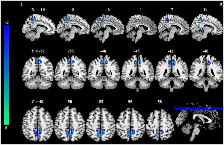 Figure 1
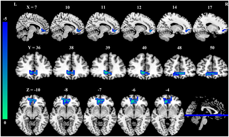 Figure 2
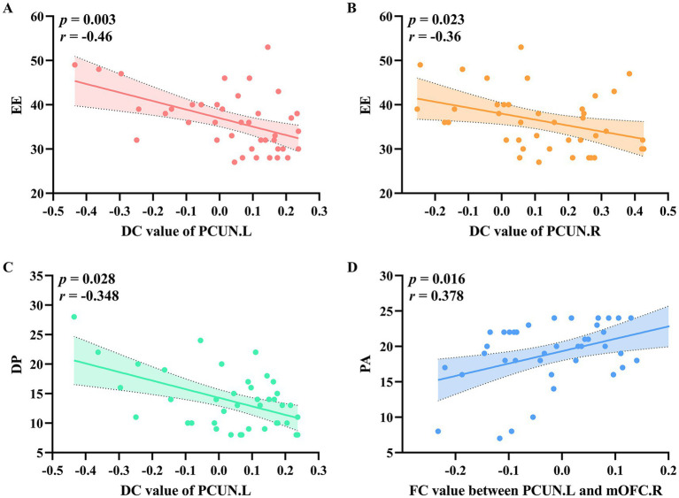 Figure 3
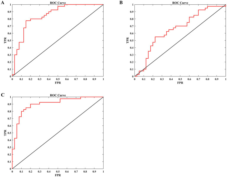 Figure 4
Peer Reviews
No public reviews on file for this paper yet. If you reviewed it on a platform where reviews are public (OpenReview, ICLR, NeurIPS, ICML), you can paste yours below so the community can read it here.
Videos
No videos yet. Explain this paper in a talk, walkthrough, or lecture? Add one.
Taxonomy
TopicsEEG and Brain-Computer Interfaces · Functional Brain Connectivity Studies