A Case of Corneal Endothelial Cell Loss After PreserFlo MicroShunt Implantation Requiring Device Removal and Ahmed Glaucoma Valve Implantation With Tube Insertion Into the Vitreous Cavity
Tomoyuki Watanabe, Koki Honzawa, Hisato Gunji, Hiroshi Horiguchi, Tadashi Nakano

TL;DR
A patient experienced significant corneal endothelial cell loss after a PreserFlo MicroShunt implant, which was resolved by removing the device and implanting an Ahmed Glaucoma Valve into the vitreous cavity.
Contribution
This case highlights a novel surgical approach using vitreous cavity tube insertion to manage complications from MicroShunt implantation.
Findings
Progressive endothelial cell loss occurred after PreserFlo MicroShunt implantation.
PMS removal and AGV implantation with vitreous cavity tube insertion stabilized IOP and endothelial cell density.
Postoperative monitoring is critical to detect and manage complications early.
Abstract
We report a case of progressive endothelial cell loss (ECL) following PreserFlo MicroShunt (PMS) (Santen Pharmaceutical, Osaka, Japan) implantation, which was successfully managed by PMS removal and the pars plana placement of Ahmed glaucoma valve (AGV) implantation. An 82-year-old man with primary open-angle glaucoma underwent PMS implantation combined with cataract surgery. Preoperative endothelial cell density (ECD) was 2,148 cells/mm², and intraocular pressure (IOP) was 21 mmHg despite five topical IOP-lowering medications. One week postoperatively, the IOP normalized to 11 mmHg without medication, but the ECD decreased to 1,400 cells/mm². Over the following 12 months, the ECD progressively declined to 880 cells/mm². PMS removal and AGV implantation, with tube insertion into the vitreous cavity, were performed to prevent further ECL and maintain IOP control. After the second…
Genes, proteins, chemicals, diseases, species, mutations and cell lines named across the full text — each resolved to its canonical identifier and authoritative record.
Click any figure to enlarge with its caption.
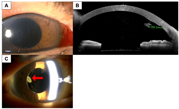 Figure 1
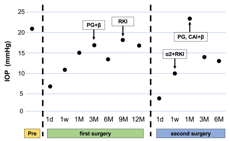 Figure 2
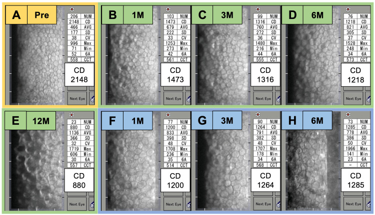 Figure 3
Peer Reviews
No public reviews on file for this paper yet. If you reviewed it on a platform where reviews are public (OpenReview, ICLR, NeurIPS, ICML), you can paste yours below so the community can read it here.
Videos
No videos yet. Explain this paper in a talk, walkthrough, or lecture? Add one.
Taxonomy
TopicsGlaucoma and retinal disorders · Corneal surgery and disorders · Retinal Imaging and Analysis