A two-step automatic identification of contrast phases for abdominal CT images based on residual networks
Qianhe Liu, Jiahui Jiang, Kewei Wu, Yan Zhang, Nan Sun, Jiawen Luo, Te Ba, Aiqing Lv, Chuane Liu, Yiyu Yin, Zhenghan Yang, Hui Xu

TL;DR
This paper introduces a two-step deep learning model using ResNet to accurately identify contrast phases in abdominal CT images, improving image quality control and AI applications.
Contribution
The novel two-step strategy using ResNet outperforms one-step methods in identifying contrast phases in abdominal CT scans.
Findings
The two-step strategy achieved 98.3% accuracy in the internal test set, significantly higher than the one-step strategy.
The two-step model reached 99.1% accuracy in the external test set with high sensitivities for all phases.
The model provides a robust tool for quality control and supports AI applications in abdominal imaging.
Abstract
To develop a deep learning model based on Residual Networks (ResNet) for the automated and accurate identification of contrast phases in abdominal CT images. A dataset of 1175 abdominal contrast-enhanced CT scans was retrospectively collected for the model development, and another independent dataset of 215 scans from five hospitals was collected for external testing. Each contrast phase was independently annotated by two radiologists. A ResNet-based model was developed to automatically classify phases into the early arterial phase (EAP) or late arterial phase (LAP), portal venous phase (PVP), and delayed phase (DP). Strategy A identified EAP or LAP, PVP, and DP in one step. Strategy B used a two-step approach: first classifying images as arterial phase (AP), PVP, and DP, then further classifying AP images into EAP or LAP. Model performance and strategy comparison were evaluated. In…
Genes, proteins, chemicals, diseases, species, mutations and cell lines named across the full text — each resolved to its canonical identifier and authoritative record.
Click any figure to enlarge with its caption.
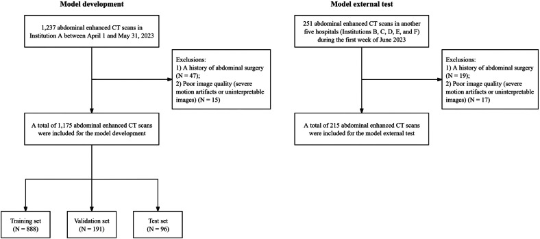 Figure 1
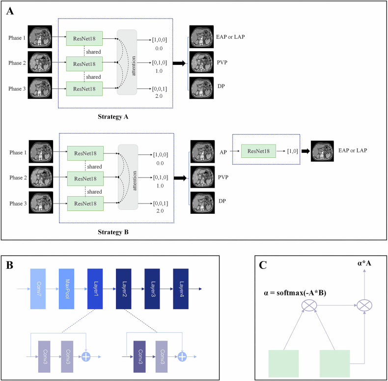 Figure 2
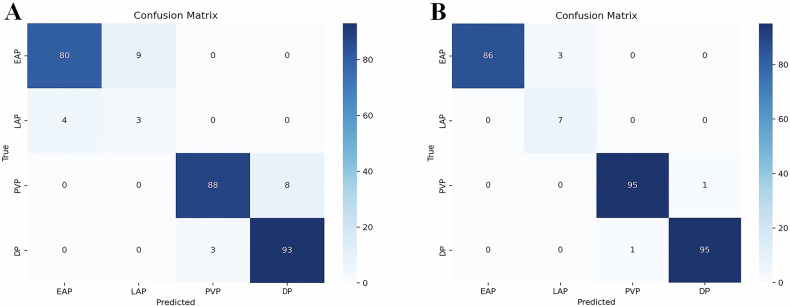 Figure 3
 Figure 4
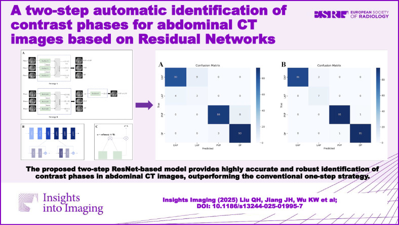 Figure 5
Peer Reviews
No public reviews on file for this paper yet. If you reviewed it on a platform where reviews are public (OpenReview, ICLR, NeurIPS, ICML), you can paste yours below so the community can read it here.
Videos
No videos yet. Explain this paper in a talk, walkthrough, or lecture? Add one.
Taxonomy
TopicsAdvanced X-ray and CT Imaging · Radiomics and Machine Learning in Medical Imaging · Radiation Dose and Imaging