7T magnetic resonance imaging-based investigation of the correlation between mammillary body structure and cognitive impairment in patients with spinocerebellar ataxia type 3
Congwei Li, Yunsong Peng, Peiling Ou, Ru Wen, Wei Chen, Chong Tian, Zhiming Zhen, Xingang Wang, Lan Ou, Chen Liu, Bijia Wang

TL;DR
This study uses 7T MRI to show that structural changes in the mammillary bodies and related brain structures are linked to cognitive impairment in patients with spinocerebellar ataxia type 3.
Contribution
The study is the first to use 7T MRI to investigate Papez circuit structures in SCA3 and their association with cognitive decline.
Findings
SCA3 patients had reduced volumes in mammillary bodies, mammillothalamic tract, and post-commissural fornix compared to healthy controls.
Cognitive impairment in SCA3 was associated with smaller left-side volumes of the mammillary bodies, mammillothalamic tract, and post-commissural fornix.
Cognitive function was positively correlated with these structures, while motor function was negatively correlated.
Abstract
Spinocerebellar ataxia type 3 (SCA3) is a hereditary disease characterized by cerebellar atrophy and motor dysfunction. Patients also exhibit non-ataxic symptoms such as cognitive impairment. While prior neuroimaging studies have identified multiple cognition-associated brain regions in SCA3 patients, research on Papez circuit structural damage (e.g., mammillary bodies (MBs)) remains sparse. Advancements in 7T magnetic resonance imaging (MRI) technology have enabled scanning and quantitative analysis of structures such as the MBs within the Papez circuit. In this study, we investigated the relationship between cognitive impairment in patients with SCA3 and structural changes in the three Papez circuit structures: the MBs, the mammillothalamic tract (MTT), and the post-commissural fornix (PF). This cross-sectional study included 46 SCA3 patients and 48 healthy controls undergoing 7T MRI…
Genes, proteins, chemicals, diseases, species, mutations and cell lines named across the full text — each resolved to its canonical identifier and authoritative record.
Click any figure to enlarge with its caption.
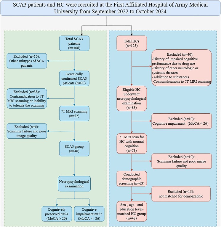 Figure 1
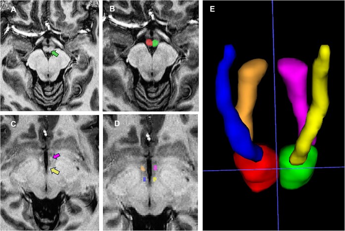 Figure 2
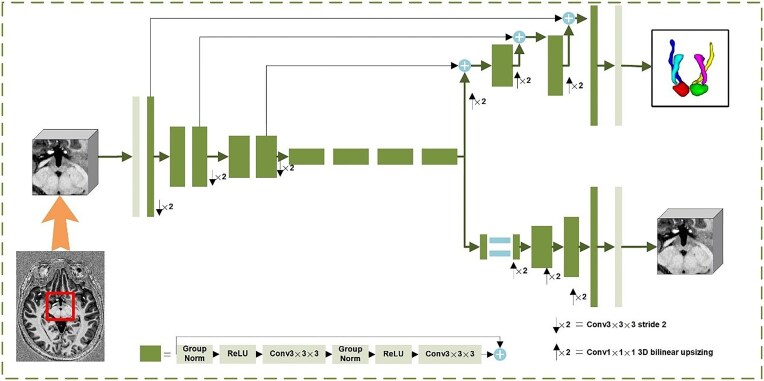 Figure 3
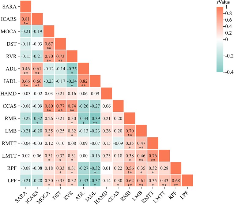 Figure 4
Peer Reviews
No public reviews on file for this paper yet. If you reviewed it on a platform where reviews are public (OpenReview, ICLR, NeurIPS, ICML), you can paste yours below so the community can read it here.
Videos
No videos yet. Explain this paper in a talk, walkthrough, or lecture? Add one.
Taxonomy
TopicsGenetic Neurodegenerative Diseases · Mitochondrial Function and Pathology · Neurological disorders and treatments