Visualizing cortical laminar architecture in the living human brain using next-generation ultra-high-gradient diffusion MRI
Susie Huang, Hansol Lee, Yixin Ma, Kwok-Shing Chan, Eva Krijnen, Laleh Eskandarian, Aneri Bhatt, Julianna Gerold, Mirsad Mahmutovic, Oula Puonti, Xiangrui Zeng, Lucas Jacob Deden Binder, Bruce Fischl, Boris Keil, Gabriel Ramos-Llordén, Eric Klawiter, Hong-Hsi Lee

TL;DR
This study uses advanced MRI to visualize the layered structure of the human brain's cortex, revealing patterns of cell and nerve density that match historical tissue samples.
Contribution
The paper introduces a non-invasive method using ultra-high-gradient diffusion MRI to map cortical laminar architecture in living humans.
Findings
Intra-soma signal fraction peaks at ~55% cortical depth, matching histological patterns.
Visual cortex has higher intra-soma signal than motor cortex in deeper layers.
Intra-soma signal correlates with cortical curvature in a layer-specific manner.
Abstract
Characterizing cortical laminar microstructure is essential for understanding human brain function. Leveraging the next-generation Connectome MRI scanner (maximum gradient strength = 500mT/m, slew rate = 600T/m/s), we characterized in vivo cortical laminar cytoarchitecture and myeloarchitecture through cortical depth-dependent analyses of soma and neurite density imaging (SANDI) metrics derived from diffusion MRI, enhanced by a super-resolution technique. SANDI revealed distinct laminar profiles: intra-soma signal fraction fis peaked at ~ 55% cortical depth, while intra-neurite signal fraction fin increased toward deeper layers, consistent with histological patterns. The visual cortex exhibited higher intra-soma signal fraction fis than the motor cortex, particularly in deeper layers. Moreover, intra-soma signal fraction fis correlated positively with cortical curvature in superficial…
Genes, proteins, chemicals, diseases, species, mutations and cell lines named across the full text — each resolved to its canonical identifier and authoritative record.
Click any figure to enlarge with its caption.
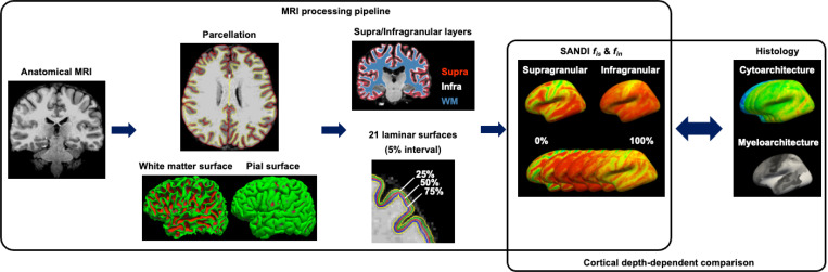 Figure 1
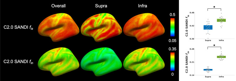 Figure 2
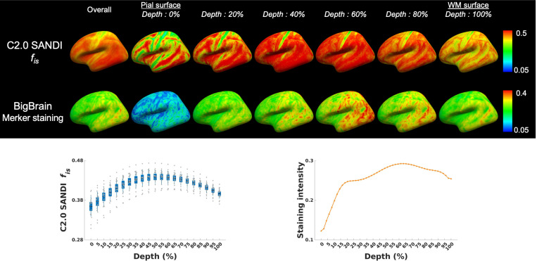 Figure 3
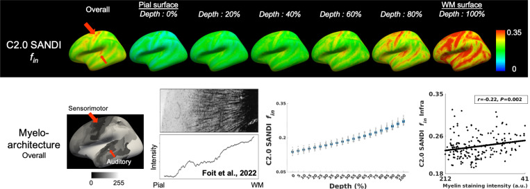 Figure 4
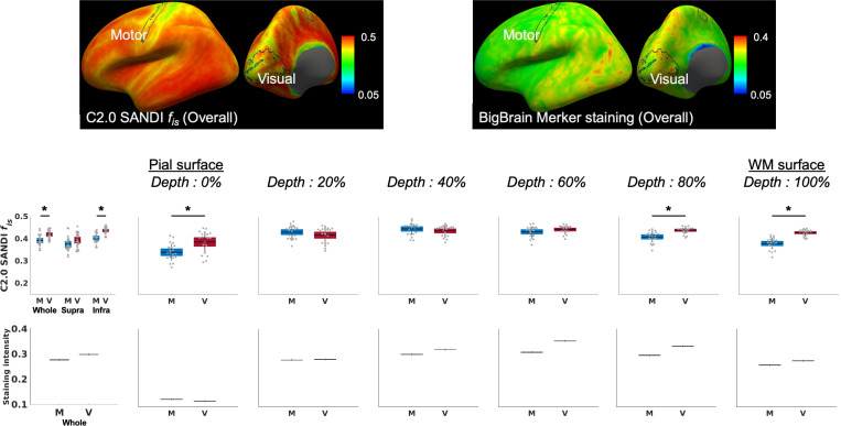 Figure 5
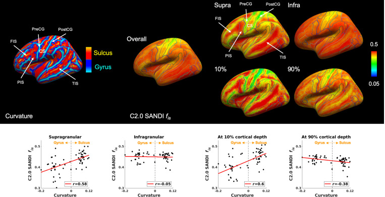 Figure 6
Peer Reviews
No public reviews on file for this paper yet. If you reviewed it on a platform where reviews are public (OpenReview, ICLR, NeurIPS, ICML), you can paste yours below so the community can read it here.
Videos
No videos yet. Explain this paper in a talk, walkthrough, or lecture? Add one.
Taxonomy
TopicsAdvanced Neuroimaging Techniques and Applications · Advanced MRI Techniques and Applications · MRI in cancer diagnosis