Meigs syndrome in low-resource setting: a pediatric case report
Sheila Macuácua, Arlindo Muhelo, Mohammad Salgado, Agostino Calção, Helton Zucula, Ahivaldino Zita, Josina Chilundo, Sandra Mavale, Damiano Pizzol, Lee Smith

TL;DR
A 13-year-old girl with late-stage Meigs Syndrome was misdiagnosed in a low-resource setting, highlighting the challenges in diagnosing this rare condition.
Contribution
This case report highlights diagnostic challenges of Meigs Syndrome in low-resource settings and emphasizes the need for awareness.
Findings
Meigs Syndrome was misdiagnosed as late-stage disease due to lack of resources.
The case underscores the importance of considering rare conditions in differential diagnosis.
Early recognition and proper diagnosis can improve outcomes in such cases.
Abstract
Meigs syndrome is a rare condition characterized by the presence of a benign fibroma of the ovary, ascites, and pleural effusion. It is very uncommon and the diagnosis is made with difficulty based on symptoms that usually mimic disseminated malignancy or tuberculosis. Although it is a benign and treatable condition, extreme presentations of late-stage diseases occur with high mortality and morbidity rates. We report on a case of a 13-year-old female presenting with misdiagnosed late-stage Meigs Syndrome in a low-resource setting.
Genes, proteins, chemicals, diseases, species, mutations and cell lines named across the full text — each resolved to its canonical identifier and authoritative record.
Click any figure to enlarge with its caption.
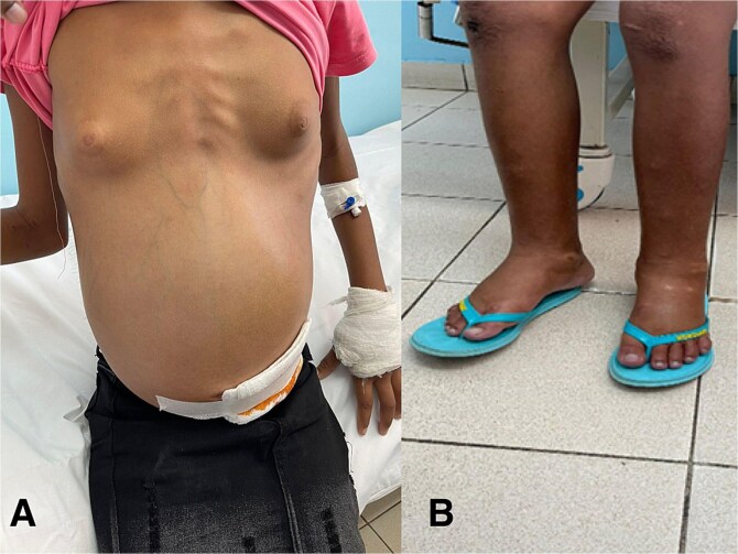 Figure 1
Peer Reviews
No public reviews on file for this paper yet. If you reviewed it on a platform where reviews are public (OpenReview, ICLR, NeurIPS, ICML), you can paste yours below so the community can read it here.
Videos
No videos yet. Explain this paper in a talk, walkthrough, or lecture? Add one.
Taxonomy
TopicsOvarian cancer diagnosis and treatment · Endometriosis Research and Treatment · Intraperitoneal and Appendiceal Malignancies
Introduction
Meigs syndrome (MS) is a rare and challenging condition characterized by the presence of benign ovarian neoplasm, ascites, and pleural effusion [1]. Only 1% of ovarian tumors present as an MS and it has been reported that 0.20 per 100 000 women are diagnosed with ovarian sex cord-stromal tumors [2]. Moreover, it is very uncommon before the third decade and the incidence progressively increases with age with a peak in the seventh decade [3]. The most common presenting symptoms are dyspnea, fatigue, and weight loss but considering the ovarian pathology, symptoms also mimic disseminated malignancy or tuberculosis [4]. The diagnosis is mainly clinical supported by imaging tests and, also some cancer markers such as carbohydrate antigen (CA) 125 and human chorionic gonadotropin (Beta-hCG) have been suggested, they are not specific nor sensitive [5]. By definition, MS is considered a benign condition treatable by exploratory laparotomy that is considered the gold standard treatment and also ascites and pleural effusion spontaneously dissolve after mass exercise [6]. In fact, although ascites and pleural effusion pathophysiological mechanisms have not been yet clarified, the most shared hypothesis is the venous and lymphatic one [7]. In presence of high-volume tumours, the partial occlusion of the venous return, leads to ascites. Then, it transudates through the capsule as the serous fluid [7]. The removal of mass may, therefore, resolve definitively these complications. Moreover, paracentesis and thoracentesis are a possible treatment for ascites and pleural effusion that can be performed to relief symptoms, mainly dyspnea [8]. In general, it has a good prognosis when early diagnosed but in low and middle-income countries extreme presentations of late-stage diseases occur with high mortality and morbidity rates mainly due to lack of diagnostic tools and training [6].
We report here a case of a 13-year-old female presenting with misdiagnosed late-stage MS in a low-resource setting.
Case report
A 13-year-old female presented with an evolution of three weeks symptomatology characterized by severe and diffuse abdominal bite-like pain type, intermittent, without periodicity with abdominal distention. After one week from symptoms onset, she was treated at the local Health Center with paracetamol and ferrous sulphate without improvement. Two weeks later the condition worsened with severe chest pain in the right hemithorax associated with dry cough and difficulty breathing. Progressively, she developed weight loss, anorexia, asthenia, postprandial infarction and edema of the lower limbs and face. She was further evaluated at the local Health Center and referred to a Central Hospital with a suspicion diagnosis of heart failure, abdominal and pulmonary tuberculosis. She presented in a moderate to severe general condition, with anasarca, pale, slight jaundice in the conjunctiva, dyspnea, apyrexia and not painful cervical lymphadenopathy. She showed decreased right thoracic expansion with 2/3 decreased vesicular murmur without adventitious noises. The abdomen was tense, with positive liquid wave sign, difficult to palpate masses, painful to superficial and deep palpation (Fig. 1A). Lower limbs were affected by grade 3 edema (Fig. 1B). A thoracentesis draining of 350 ml of citrus yellow fluid was performed and resulted negative for culture, cytochemical and genexpert tests and neoplastic cell research. A paracentesis draining of 500 ml of milky ascitic liquid was performed and resulted in relief of respiratory distress and abdominal pain. An ultrasound showed bilateral pleural effusion, mainly on the right and pelvic bilateral masses. Cancer markers were negative for Alpha-1phetoprotein (1.31 U/ml) and carcinoembryonic antigen (CEA) (0.76 U/ml) and positive for CA 125 (144.5 U/ml) and Beta-hCG (2.12 U/ml). The patient was therefore admitted to the Gynecology Department. During the hospitalization she was treated with ceftriaxone and vancomycin, and a transfusion of red blood cell concentrate was performed. After 2 days her clinical conditions worsened, with significant respiratory distress, followed by cardiorespiratory arrest and death. The autopsy examination and histologic exam confirmed the presence of fibromas on both ovaries.
Pediatric Meigs syndrome at presentation: Tense abdomen (A) and lower limbs edema (B).
Discussion
We reported a rare and extraordinary case of pediatric MS that reflects the poor social-economic context and limited resources of the context in which health care workers must work in low-resource settings. MS is, per se, a rare condition but in this case, it was even more extraordinary considering that the syndrome mainly affects women over the age of 30 while the patient was just 13 years old [9]. Proper diagnosis and management are of paramount importance for all conditions and for MS as it is considered a benign and treatable condition when treated properly and on time [10]. In very young patients as our case, CA 125 and Beta-hCG markers can play an important role in diagnostic flow, although they are not specific for this disease. Due to economic issues and transportation difficulties, the patient arrived at the referral hospital after three weeks of symptoms onset and in an already compromised general status. This initial inadequate treatment and delay in transfer demonstrates the limited resources and equipment of rural facilities and the lack of proper training and ability to recognize such an emergency. Once she presented, she was already in a critical condition, and there was limited time to stabilize her and to perform a laparotomy. There was only time to perform thoracentesis and paracentesis and relief of symptoms, but not to reverse general conditions which led to her death.
This case highlights the fragility of low-income healthcare systems mainly due to the lack of specialized and well trained health workers and equipment. For this, it is mandatory to develop effective public health policies that address these disparities and provide enhanced protection for vulnerable patients. In particular, it is crucial to promote early diagnosis both promoting patient care accessibility, both improving health workers access to diagnostic tools. In low-resource settings also telemedicine could play a crucial role in detection and management of rare conditions like S with further positive impact on all treatable diseases, especially for most vulnerable population.
The reference list from the paper itself. Each links out to its DOI / PubMed record.
- 1Iavarone I, Padovano M, Pasanisi F. et al. Meigs syndrome and elevated CA-125: case report and literature review of an unusual presentation mimicking ovarian cancer. Med (Kaunas) 2023;59:1684. 10.3390/medicina 59091684 PMC 1053583037763803 · doi ↗ · pubmed ↗
- 2Riker D, Goba D. Ovarian mass, pleural effusion, and ascites: revisiting Meigs syndrome. J Bronchology Interv Pulmonol 2013;20:48–51. 10.1097/LBR.0b 013e 31827 ccb 3523328144 · doi ↗ · pubmed ↗
- 3Mohammed SA, Kumar A. Meigs Syndrome. 2023 Jul 4. In: Stat Pearls [Internet]. Stat Pearls Publishing: Treasure Island (FL), 2024.
- 4Antunes M, Pizzol D, Zambon M. et al. Giant ovarian fibroma with associated Meigs syndrome in low resources setting. J Surg Case Rep 2019;2019:rjz 143. 10.1093/jscr/rjz 143PMC 658240431231501 · doi ↗ · pubmed ↗
- 5Sofoudis C, Kouiroukidou P, Louis K. et al. Enormous ovarian fibroma with elevated Ca-125 associated with Meigs' syndrome. Presentation of a rare case. Eur J Gynaecol Oncol 2016;37:142–3.27048129 · pubmed ↗
- 6Ali AA, Jeannot BM, Olasinde AA. et al. A rare case of demons-Meigs' syndrome with a 7.5 kg giant ovarian fibroma associated with severe dyspnea: case report. Ann Med Surg (Lond) 2023;85:6243–6. 10.1097/MS 9.000000000000142438098603 PMC 10718357 · doi ↗ · pubmed ↗
- 7Dockerty MB, Masson JC. Ovarian fibromas: a clinical and pathologic study of two hundred and eighty-three cases. Am J Obstet Gynecol 1944;47:741–52. 10.1016/S 0002-9378(16)40377-7 · doi ↗
- 8Bahall V, De Barry L, Singh K. Thoracic endometriosis masquerading as Meigs' syndrome in a young woman: a case report and literature review. Case Rep Womens Health 2022;36:e 00452. 10.1016/j.crwh.2022.e 0045236246455 PMC 9562932 · doi ↗ · pubmed ↗