Intraductal ultrasonography using an ultrathin radial miniature probe and guide sheath to diagnose a bile duct stricture
Tsuyoshi Suda, Kiichiro Kaji, Miyabi Miura, Kuniaki Arai, Shuichi Terasaki

Abstract
Genes, proteins, chemicals, diseases, species, mutations and cell lines named across the full text — each resolved to its canonical identifier and authoritative record.
Click any figure to enlarge with its caption.
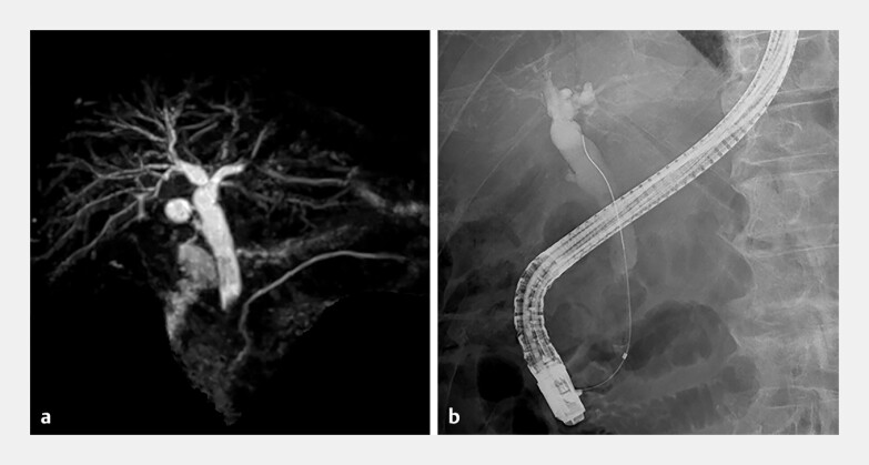 Fig. 1
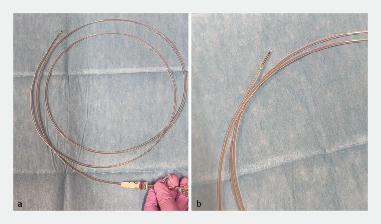 Fig. 2
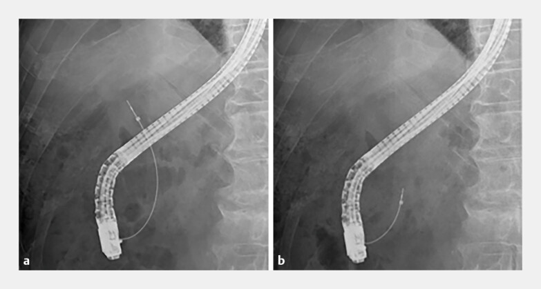 Fig. 3
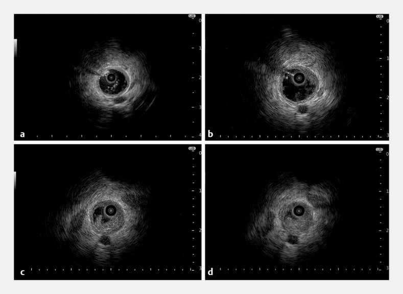 Fig. 4
Peer Reviews
No public reviews on file for this paper yet. If you reviewed it on a platform where reviews are public (OpenReview, ICLR, NeurIPS, ICML), you can paste yours below so the community can read it here.
Videos
No videos yet. Explain this paper in a talk, walkthrough, or lecture? Add one.
Taxonomy
TopicsPediatric Hepatobiliary Diseases and Treatments · Gallbladder and Bile Duct Disorders · Gastrointestinal disorders and treatments
A 91-year-old man presented with liver dysfunction: aspartate aminotransferase 81 U/L, alanine aminotransferase 166 U/L, alkaline phosphatase 323 U/L, gamma-glutamyl transferase 527 U/L, total bilirubin 1.6 mg/dL, prothrombin time-international normalized ratio 1.02, and albumin 3.5 g/dL. Magnetic resonance cholangiopancreatography and endoscopic retrograde cholangiopancreatography (ERCP) revealed a distal bile duct stricture ( Fig. 1 ). Laboratory tests revealed an elevated C-reactive protein level (4.30 mg/dL), and acute cholangitis was suspected. After endoscopic biliary drainage had been performed, adenocarcinoma was identified from bile cytology. Contrast-enhanced computed tomography failed to clearly delineate the tumor.
A lower bile duct stricture is shown on: a magnetic resonance cholangiopancreatography: b endoscopic retrograde cholangiopancreatography.
Further diagnostic evaluation was conducted using ERCP and intraductal ultrasonography (IDUS). An ultrathin radial miniature probe (UM-S20-17S; Olympus, Tokyo, Japan) with a 1.4-mm distal diameter was deployed through a guide sheath system (UMIDAS sheath cannula; UMIDAS, Kanagawa, Japan) ( Fig. 2 ; Video 1 ), as confirmed on fluoroscopic images ( Fig. 3 ). Detailed IDUS at 20 MHz, performed while infusing saline via the guide sheath to distend the bile duct, showed normal proximal bile ducts ( Fig. 4 a ), the tumor’s upper margin ( Fig. 4 b ), the stricture site ( Fig. 4 c ), and serosal layer disruption ( Fig. 4 d ). After the IDUS, forceps biopsies were obtained through the guide sheath, which confirmed the diagnosis of adenocarcinoma. The patient opted for best supportive care.
a, b Photographs of the ultrathin radial miniature probe being passed through a guide sheath system.
a, b Fluoroscopic images showing the ultrathin radial miniature probe passing through the guide sheath system into the bile duct.
Detailed intraductal ultrasonography images showing: a normal proximal bile ducts; b the upper margin of the tumor; c the stricture site; d disruption of the serosal layer.
Ultrasonographic observation of the bile duct using an ultrathin radial miniature probe passed through a guide sheath, performed while infusing saline solution.Video 1
Radial miniature probes, such as the UM-S20-17S, are primarily used in respiratory medicine for endobronchial ultrasound 1 . Despite its potential advantages, the extreme thinness of the UM-S20-17S may explain its limited use during ERCP at many facilities. The guide sheath system was originally developed for selective pancreatic and biliary duct biopsy 2 , but other applications have been reported, including those previously documented by our group 3 4 . IDUS uses a high frequency ultrasonography probe to obtain real-time high quality cross-sectional images during ERCP; however, the challenge of maintaining the probe at the center of the bile duct and the presence of air within the duct can affect image quality, making it difficult to produce clear images 5 . Observing the bile duct using an ultrathin radial miniature probe passes through a guide sheath may address these issues.
Endoscopy_UCTN_Code_TTT_1AR_2AD
The reference list from the paper itself. Each links out to its DOI / PubMed record.
- 1Tanaka M Matsumoto Y Imabayashi T Diagnostic value of a new cryoprobe for peripheral pulmonary lesions: a prospective study BMC Pulm Med 20222222610.1186/s 12890-022-02003-035689261 PMC 9188163 · doi ↗ · pubmed ↗
- 2Kawakami H Uchiyama N Hatada H Newly developed dedicated guide sheath system for selective pancreatobiliary biopsy Dig Endosc 202335 e 100e 10210.1111/den.1459337246406 · doi ↗ · pubmed ↗
- 3Miwa H Oishi R Endo K Antegrade metallic stent placement using a slim cholangioscope for malignant afferent loop obstruction Endoscopy 202456 E 774E 77510.1055/a-2387-423839231522 PMC 11374441 · doi ↗ · pubmed ↗
- 4Suda T Satomura K Miura M Endoscopic removal of a deteriorated fully-covered self-expandable metal stent using an endoscopic retrograde cholangiopancreatography guide sheath Clin Endosc 20255814714810.5946/ce.2024.17039722142 PMC 11837560 · doi ↗ · pubmed ↗
- 5Sun B Hu B The role of intraductal ultrasonography in pancreatobiliary diseases Endosc Ultrasound 2016529129910.4103/2303-9027.19160727803901 PMC 5070286 · doi ↗ · pubmed ↗