Peroral direct cholangioscopy and snare technique for stray bile duct stone for patients who have undergone bile duct jejunostomy
Nobuhiko Fukuba, Hiroyuki Fukuhara, Yoshiko Takahashi, Yasuhide Kodama, Masaki Onoe, Shuichi Sato, Shunji Ishihara

Abstract
Genes, proteins, chemicals, diseases, species, mutations and cell lines named across the full text — each resolved to its canonical identifier and authoritative record.
Click any figure to enlarge with its caption.
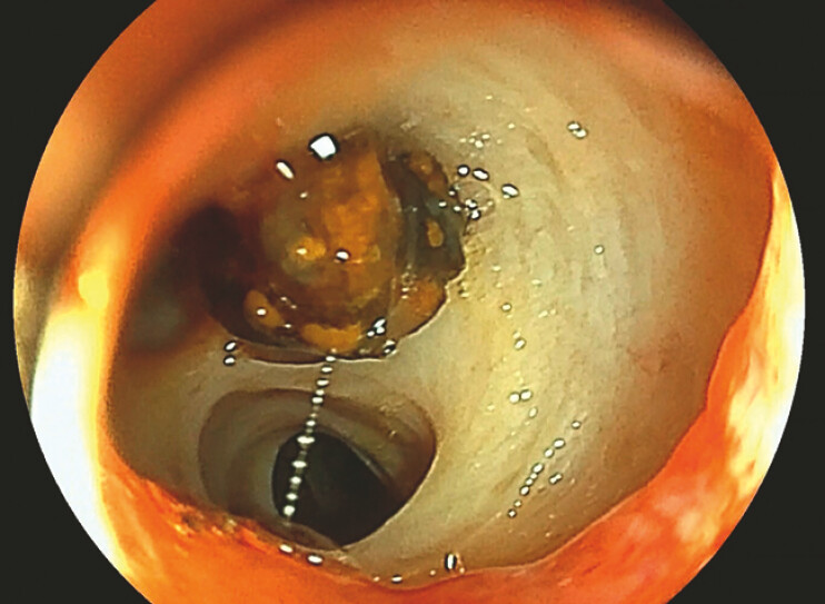 Fig. 1
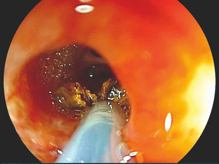 Fig. 2
Peer Reviews
No public reviews on file for this paper yet. If you reviewed it on a platform where reviews are public (OpenReview, ICLR, NeurIPS, ICML), you can paste yours below so the community can read it here.
Videos
No videos yet. Explain this paper in a talk, walkthrough, or lecture? Add one.
Taxonomy
TopicsGallbladder and Bile Duct Disorders · Biliary and Gastrointestinal Fistulas · Esophageal and GI Pathology
A male patient over 80 years old, who underwent a pancreaticoduodenectomy 7 years prior, was presented with transient abdominal pain. Computed tomography (CT) findings indicated the presence of a common bile duct stone, thus a balloon-assisted enteroscopy-assisted endoscopic retrograde cholangiopancreatography (BAE-ERCP) procedure was performed. However, the stone could not be identified in the cholangiography images due to gas reflux, while sweeping with a basket was also ineffective.
After 2 months, the patient experienced abdominal pain recurrence and subsequent CT scanning revealed the previously identified stone. It was determined that BAE-ERCP alone would not be able to identify the stone, thus a peroral direct cholangioscopy (PDCS) examination was performed. The procedure was initiated with the insertion of a double-balloon endoscope (EI-580BT, Fujifilm, Tokyo) into the area of the previous bile duct jejunal anastomosis, followed by its removal with the overtube remaining ( Video 1 ). Next, balloon dilation was performed, and a slit was made in the overtube, through which a small-caliber endoscope (SCE) (EG-740N, Fujifilm, Tokyo) was inserted. The SCE was then fitted tightly into the anastomosis and observations of the interior of the bile duct revealed that the stone had become trapped inside during the suction procedure, similar to a ball-check valve ( Fig. 1 ). The stone was grasped using a snare (Snare Master, Olympus), then crushed and removed by aspiration with the SCE ( Fig. 2 ).
The stone had become trapped within the anastomosis, similar to a ball-check valve.
The stone was easily grasped with the snare and split into two pieces with little force required.
During the anastomosis procedure, observation of the internal portion of the bile duct with the SCE revealed a trapped stone, similar to a ball-check valve. A snare (Snare, Fujifilm) was used to grasp the stone, allowing it to be crushed and then removed by aspiration with the SCE.Video 1
The effectiveness of BAE-ERCP for cases with a reconstructed intestine has been reported, though stone removal is generally difficult 1 2 3 4 . The SCE used has a 2.4-mm channel diameter, which facilitated the utilization of a snare and allowed the stone pieces to be removed by suction. It is difficult to identify floating stones in bile duct jejunal anastomosis cases using cholangiography, due to gas reflux. PDCS was found to be effective in the present case in identifying and efficiently removing the stone.
Endoscopy_UCTN_Code_TTT_1AP_2AD
The reference list from the paper itself. Each links out to its DOI / PubMed record.
- 1Matsushita M Shimatani M Ikeura T Peroral direct cholangioscopy with an ultraslim gastroscope in combination with a short double-balloon enteroscope in patients with altered GI anatomy Gastrointest Endosc 20107188420363436 10.1016/j.gie.2009.08.004 · doi ↗ · pubmed ↗
- 2Matsushita M Shimatani M Ikeura T Peroral direct cholangioscopy with an ultraslim gastroscope in combination with a short double-balloon enteroscope for reconstructed biliary anatomy Endoscopy 201143101722057770 10.1055/s-0030-1256698 · doi ↗ · pubmed ↗
- 3Itoi T Sofuni A Itokawa F Diagnostic and therapeutic peroral direct cholangioscopy in patients with altered GI anatomy (with videos)Gastrointest Endosc 20127544144922154415 10.1016/j.gie.2011.09.038 · doi ↗ · pubmed ↗
- 4Sola-Vera J Uceda F Cuesta R Direct peroral cholangioscopy using an ultrathin endoscope: making technique easier Revista Española de Enfermedades Digestivas 2014106303610.4321/s 1130-0108201400010000524689713 · doi ↗ · pubmed ↗