Endoscopic drainage for the treatment of intramural colonic abscess
Yuki Amioka, Satoshi Masuda, Hidenori Tanaka, Ken Yamashita, Yoshihiro Kishida, Toshio Kuwai, Shiro Oka

Abstract
Genes, proteins, chemicals, diseases, species, mutations and cell lines named across the full text — each resolved to its canonical identifier and authoritative record.
Click any figure to enlarge with its caption.
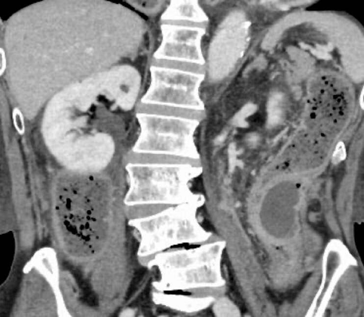 Fig. 1
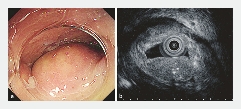 Fig. 2
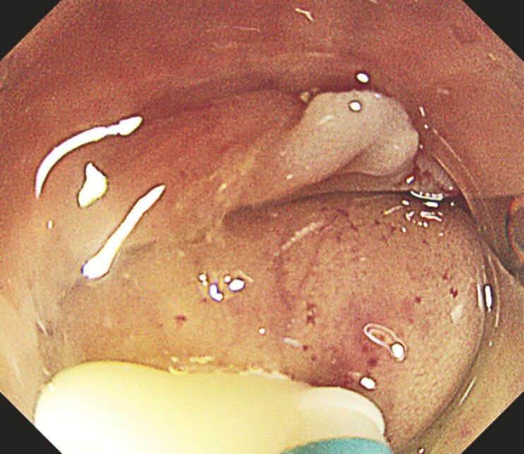 Fig. 3
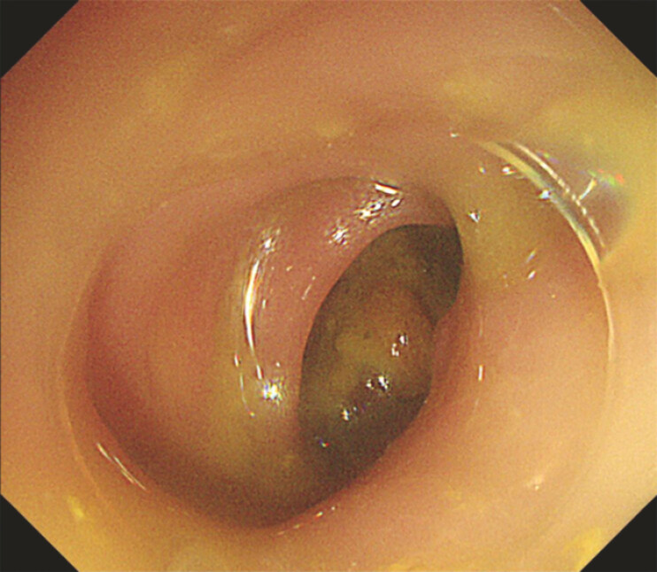 Fig. 4
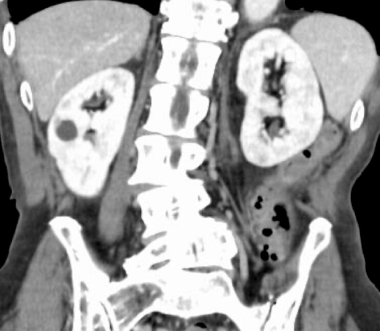 Fig. 5
Peer Reviews
No public reviews on file for this paper yet. If you reviewed it on a platform where reviews are public (OpenReview, ICLR, NeurIPS, ICML), you can paste yours below so the community can read it here.
Videos
No videos yet. Explain this paper in a talk, walkthrough, or lecture? Add one.
Taxonomy
TopicsGastrointestinal disorders and treatments · Esophageal and GI Pathology · Diverticular Disease and Complications
A 70-year-old man presented to our hospital with abdominal pain and vomiting. Contrast-enhanced computed tomography (CT) revealed a capsulated abscess adjacent to the descending colon causing intestinal obstruction ( Fig. 1 ). A retrospective review of a CT scan from 2 years prior identified a linear hyperdense structure resembling a fish bone penetrating the intestinal wall at the same site, which was suspected to be the cause of the abscess.
A capsulated abscess causing intestinal obstruction.
Colonoscopy revealed a submucosal tumor-like elevation corresponding to the abscess ( Fig. 2 a ), with luminal obstruction preventing the passage of the scope. Endoscopic ultrasonography (EUS) demonstrated a homogeneous hypoechoic lesion with internal hyperechoic foci, predominantly in the submucosa, with a preserved muscularis propria ( Fig. 2
b ). Given the intramural nature of the abscess, percutaneous CT-guided drainage posed a risk of perforation or fistula formation. Therefore, endoscopic drainage was attempted via colonoscopy ( Video 1 ).
a Colonoscopy revealed a submucosal tumor-like elevation. b Endoscopic ultrasonography demonstrated a homogeneous hypoechoic lesion with internal hyperechoic foci.
By utilizing EUS and CT for precise diagnosis, we safely performed minimally invasive endoscopic drainage for the treatment of intramural colonic abscess, achieving symptom resolution.Video 1
A 10 mm mucosal incision was made using DualKnife J (Olympus Medical Systems), leading to purulent evacuation ( Fig. 3 ). Sufficient drainage via suction and mucosal compression allowed the scope to pass ( Fig. 4 ). The symptoms resolved immediately, and the patient was discharged 9 days later. Follow-up CT 2 months later confirmed the resolution of the abscess with minor residual intramural air ( Fig. 5 ).
A 10 mm mucosal incision was made using DualKnife J, leading to purulent evacuation.
Sufficient drainage via suction and mucosal compression allowed the scope to pass.
Follow-up CT 2 months later confirmed the resolution of the abscess with minor residual intramural air. Abbreviation: CT, computed tomography.
Colonic intramural abscesses are rare, and to our knowledge, there have been no previous reports of endoscopic incision and drainage for relief of obstruction, although several cases have been reported in the esophagus and stomach 1 2 3 . Recently, EUS-guided drainage has proven useful for extramural abdominal abscesses 4 . In this case of intramural abscess, we performed minimally invasive endoscopic drainage via mucosal incision alone. Procedural safety was ensured through precise diagnosis with EUS and CT. This case highlights the potential of endoscopic treatment as a viable alternative to surgical or percutaneous drainage for colonic intramural abscesses.
Endoscopy_UCTN_Code_TTT_1AS_2AJ
The reference list from the paper itself. Each links out to its DOI / PubMed record.
- 1Jiyu Z Huige W Dan L Treatment of a spontaneous intramural esophageal abscess by endoscopic mucosal incision: an easy and effective therapy Endoscopy 202254 E 937E 93835835156 10.1055/a-1882-5422 PMC 9736806 · doi ↗ · pubmed ↗
- 2Kim KR Shim KN Choe ARA case of intramural gastric wall abscess, a rare disease successfully treated with endoscopic incision and drainage Gut Liver 20231794995336700301 10.5009/gnl 220009 PMC 10651380 · doi ↗ · pubmed ↗
- 3Takeno S Moroga T Ono K Endoscopic mucosal incision for successful treatment of submucosal abscess extending the full length of the esophagus due to fish bone: report of a cese Esophagus 201512199202
- 4Liu S Tian Z Jiang Y Endoscopic ultrasoundguided drainage to abdominal abscess: A systematic review and metaanalysis J Min Access Surg 20221848949610.4103/jmas.jmas_349_21PMC 963269535915535 · doi ↗ · pubmed ↗