The guidewire-guided endoscopic retrograde direct cholangioscopy facilitating the difficult biliary cannulation of small papilla
Shan-Shan Hu, Xiao-Gang Liu, Jie Hou, Wei-Hui Liu

Abstract
Genes, proteins, chemicals, diseases, species, mutations and cell lines named across the full text — each resolved to its canonical identifier and authoritative record.
Click any figure to enlarge with its caption.
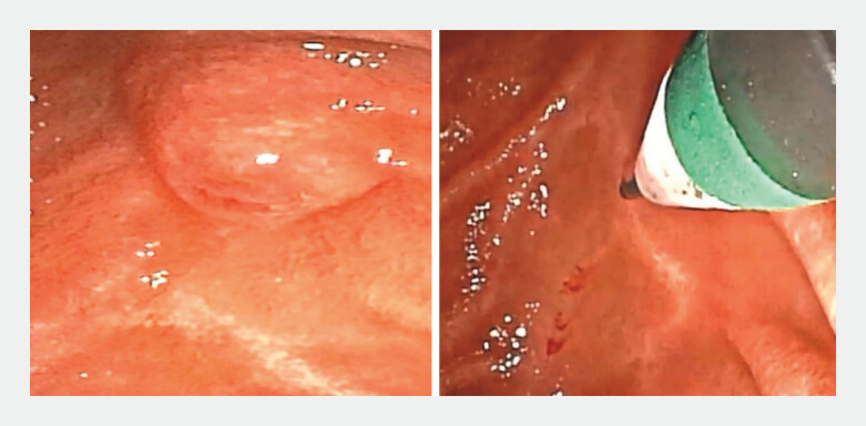 Fig. 1
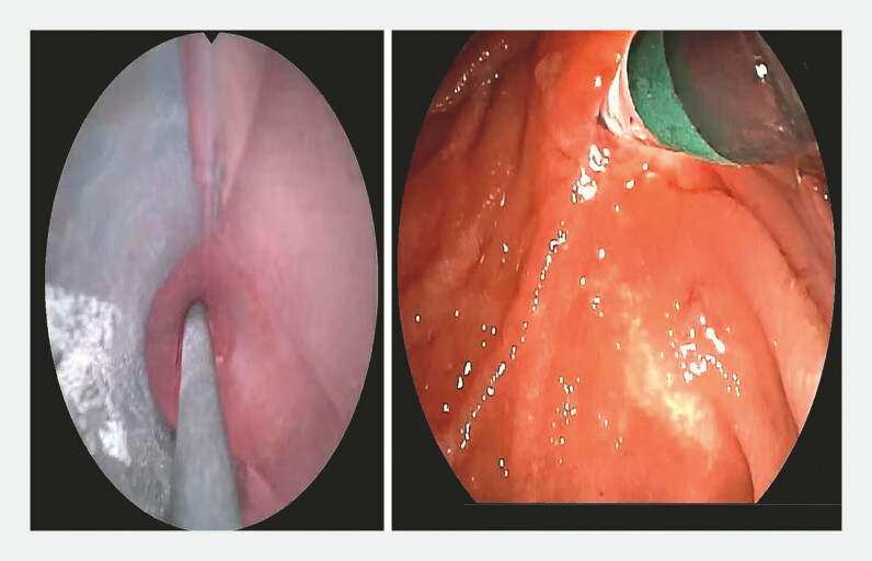 Fig. 2
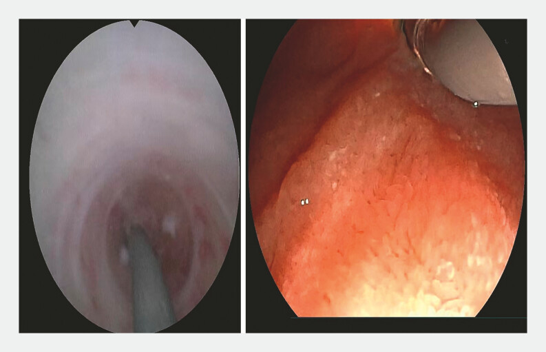 Fig. 3
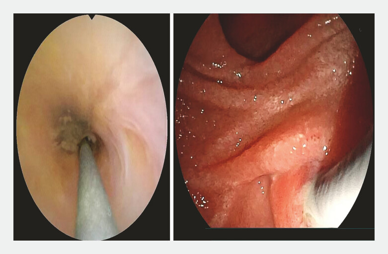 Fig. 4
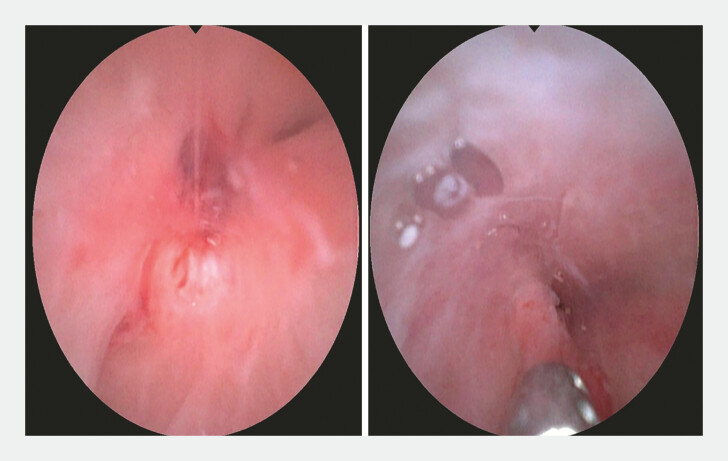 Fig. 5
- —Department of Science & Technology Department of Sichuan Province
- —The Program for Sichuan Medical and Health Care Promotion Institute
Peer Reviews
No public reviews on file for this paper yet. If you reviewed it on a platform where reviews are public (OpenReview, ICLR, NeurIPS, ICML), you can paste yours below so the community can read it here.
Videos
No videos yet. Explain this paper in a talk, walkthrough, or lecture? Add one.
Taxonomy
TopicsGallbladder and Bile Duct Disorders · Pediatric Hepatobiliary Diseases and Treatments · Esophageal and GI Pathology
Our team successfully developed a novel endoscopic retrograde direct cholangioscopy (ERDC) technique, which involves attaching a conical transparent cap to the front end of the cholangioscope, enabling intubation under direct vision 1 2 . However, in clinical practice, we observed variations in intubation success rates depending on papillary morphology 3 . For smaller, more challenging papillae, we learned from the traditional endoscopic retrograde cholangiopancreatography (ERCP) intubation method 4 , introducing a wire-guided ERDC strategy that significantly improves success rates for small papillae ( Video 1 ).
The guidewire-guided endoscopic retrograde direct cholangioscopy facilitating the difficult biliary cannulation of small papillaVideo 1
A male patient presented with an unexplained thickening wall of the common bile duct (CBD). ERDC was planned for direct visualization and comprehensive examination of the CBD. During duodenoscopy, the papillary orifice was found too small, preventing the conical transparent cap at the distal end of the choledochoscope from entering the papilla, resulting in failed ERDC intubation. To overcome this, we extended the guidewire tip approximately 2 mm beyond the conical transparent cap ( Fig. 1 ), inserted it into the papillary opening along the CBD’s direction, and then slipped into the CBD ( Fig. 2 ). With the guidance of guidewire, the choledochoscope able to be inserted into the small papilla ( Fig. 3 ). When the guidewire was inserted forward, the yellow bile was observed under direct vision, thus cholangioscope could be cannulated into the CBD ( Fig. 4 ). This approach successfully enabled full visualization and biopsy sampling of the distalCBD ( Fig. 5 ).
A conical transparent cap was attached to the distal end of the choledochoscope, with the guidewire tip extending approximately 2 mm beyond the cap.
The guidewire was first inserted into the papillary orifice.
The guidewire was advanced into the common bile duct.
The guidewire was manipulated back and forth, allowing bile to flow out, confirming entry into the bile duct.
The choledochoscope was smoothly advanced along the guidewire into the common bile duct, enabling full visualization and biopsy at the lesion site.
For cases with a small papilla or when the choledochoscope's distal end exceeds the papillary orifice, this method is particularly useful. The guidewire plays a crucial guiding role, potentially improving the success rate of difficult ERDC cannulation in the future.
Endoscopy_UCTN_Code_TTT_1AR_2AB
The reference list from the paper itself. Each links out to its DOI / PubMed record.
- 1Liu WH Huang XY Hu X Initial experience of visualized biliary cannulation during ERCP Endoscopy 2023551037104210.1055/a-2113-895237339664 · doi ↗ · pubmed ↗
- 2Liu WH Huang XY Zhang RY From darkness to brightness: the cholangioscopy-guided selective biliary cannulation with the help of transparent cap during ERCP Endoscopy 20235501 E 320E 32110.1055/a-1981-250336513111 PMC 9833945 · doi ↗ · pubmed ↗
- 3Haraldsson E Lundell L Swahn F Endoscopic classification of the papilla of Vater. Results of an inter- and intraobserver agreement study United European Gastroenterol J 2017550451010.1177/2050640616674837 PMC 544615028588881 · doi ↗ · pubmed ↗
- 4Testoni PA Mariani A Aabakken L Papillary cannulation and sphincterotomy techniques at ERCP: European Society of Gastrointestinal Endoscopy (ESGE) Clinical Guideline Endoscopy 20164865768310.1055/s-0042-10864127299638 · doi ↗ · pubmed ↗