Value of preoperative enteroscopic carbon nanoparticle labeling in guiding laparoscopic resection of Meckel’s diverticulum
Chen Wu, Lingyun Wang, Xiangyu Liu, Chunlin Wang, Jing Cao

Abstract
Genes, proteins, chemicals, diseases, species, mutations and cell lines named across the full text — each resolved to its canonical identifier and authoritative record.
Click any figure to enlarge with its caption.
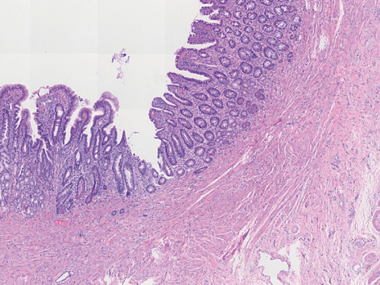 Fig. 1
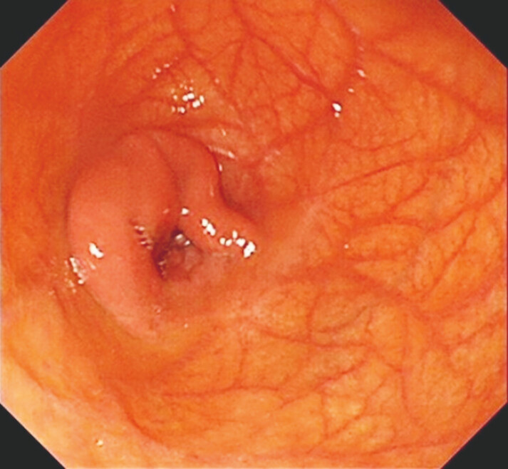 Fig. 2
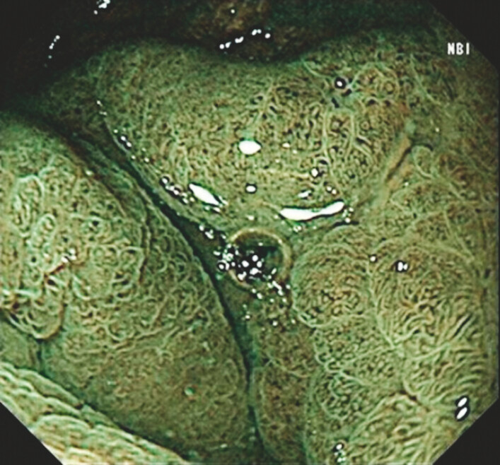 Fig. 3
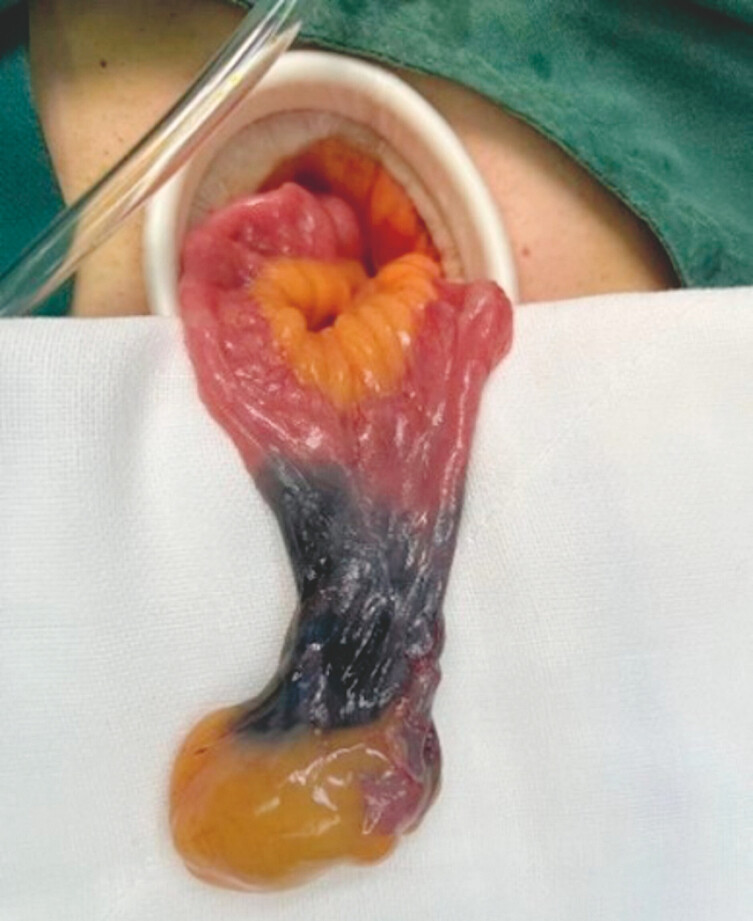 Fig. 4
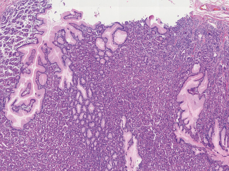 Fig. 5
Peer Reviews
No public reviews on file for this paper yet. If you reviewed it on a platform where reviews are public (OpenReview, ICLR, NeurIPS, ICML), you can paste yours below so the community can read it here.
Videos
No videos yet. Explain this paper in a talk, walkthrough, or lecture? Add one.
Taxonomy
TopicsGastrointestinal disorders and treatments · Gastrointestinal Tumor Research and Treatment · Esophageal and GI Pathology
Meckel’s diverticulum is a congenital digestive tract malformation, with a prevalence of 0.3% to 2.9% in the general population 1 2 3 . Only 15% of patients with Meckel’s diverticulum are symptomatic and preoperative diagnosis with gastrointestinal endoscopy is limited 3 . Enteroscopy provides precise anatomical guidance for minimally invasive surgery. Herein, we report two rare cases of patients presenting with hematochezia. In both cases, enteroscopy revealed the Meckel’s diverticulum, and carbon nanoparticle labeling was subsequently performed. The lesions were resected via laparoscopic surgery guided by carbon nanoparticle labeling ( Video 1 ).
Value of preoperative enteroscopic carbon nanoparticle labeling in guiding laparoscopic resection of Meckel’s diverticulum.Video 1
Patient 1: A 20-year-old man presented with a 4-day history of hematochezia. Previous gastrointestinal endoscopy failed to identify the bleeding source. Subsequent enteroscopy revealed a diverticulum with a narrow opening, located 200 cm proximal to the ileocecal valve. Submucosal injection of carbon nanoparticles was performed around the lesion for marking. Then, laparoscopic exploration guided by carbon nanoparticle labeling confirmed the diverticulum, measuring 4 × 1.2 cm, and segmental small-bowel resection was performed. Histopathology confirmed Meckel’s diverticulum with normal ileal wall ( Fig. 1 ).
Microscopic appearance. A loupe view of the resected Meckel’s diverticulum showed normal ileal wall with well-formed villi and the muscularis propria (hematoxylin and eosin ×4).
Patient 2: A 17-year-old man was admitted with recurrent hematochezia for over 1 year and recurrence for 3 days. Previous colonoscopy failed to identify the bleeding source. Subsequent enteroscopy found a giant diverticulum with the blind end of the diverticulum showing irregular mucosal protrusions ( Fig. 2 ). Narrow-band imaging showed heterotopic gastric mucosa characteristics ( Fig. 3 ). A tortuous submucosal artery with visible pulsation was observed adjacent to the lesion. The lesion was marked using carbon nanoparticles and a preliminary diagnosis of Meckel’s diverticulum was established. Then, the patient was transferred for laparoscopic surgical intervention. Laparoscopic exploration revealed the giant diverticulum in the distal ileum, approximately 100 cm proximal to the ileocecal valve ( Fig. 4 ). Histopathology confirmed Meckel’s diverticulum containing heterotopic gastric mucosa ( Fig. 5 ).
The enteroscopy found a giant diverticulum with the blind end of the diverticulum showing irregular mucosal protrusions.
Narrow-band imaging showed heterotopic gastric mucosa characteristics.
Laparoscopic surgery revealed Meckel’s diverticulum in the ileum, approximately 100 cm proximal to the ileocecal valve.
Microscopic findings. A loupe view of the heterotopic gastric mucosa of Meckel’s diverticulum (hematoxylin and eosin ×4).
Enteroscopy with carbon nanoparticle labeling provides precise anatomical guidance for minimally invasive surgery.
Endoscopy_UCTN_Code_CCL_1AC_2AF
The reference list from the paper itself. Each links out to its DOI / PubMed record.
- 1Bejiga G Ahmed Z Gangrenous Meckel’s diverticulum with small bowel obstruction mimicking complicated appendicitis: ‘case report’Int J Surg Case Rep 20229710741910.1016/j.ijscr.2022.107419 PMC 940320135863288 · doi ↗ · pubmed ↗
- 2Hu S Du H Wen J Diagnosis of inverted Meckel’s diverticulum by double-balloon enteroscopy: a case report AME Case Rep 202483310.21037/acr-23-10238711899 PMC 11071006 · doi ↗ · pubmed ↗
- 3Gomes GF Bonin EA Noda RW Balloon-assisted enteroscopy for suspected Meckel’s diverticulum and indefinite diagnostic imaging workup World J Gastrointest Endosc 2016867968310.4253/wjge.v 8.i 18.67927803776 PMC 5067476 · doi ↗ · pubmed ↗