Guidewire-assisted endoscopic mucosal resection with over-the-scope clips for a cecal submucosal tumor
Toru Kuwano, Shunji Shimaoka, Hirotake Kusumoto, Hideyuki Kishita, Tsutomu Sakiyama, Yukiko Baba, Saori Furukawa

Abstract
Genes, proteins, chemicals, diseases, species, mutations and cell lines named across the full text — each resolved to its canonical identifier and authoritative record.
Click any figure to enlarge with its caption.
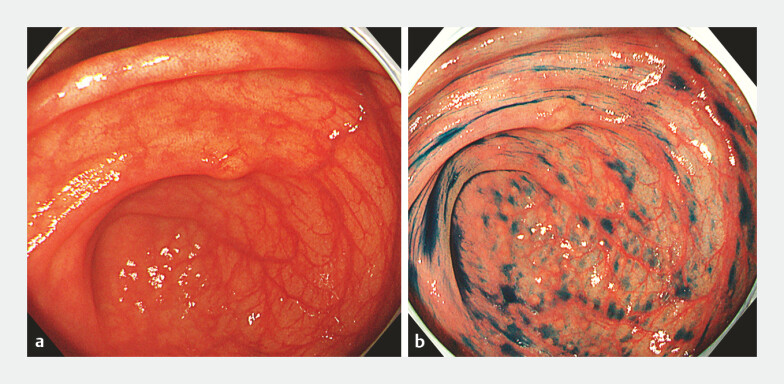 Fig. 1
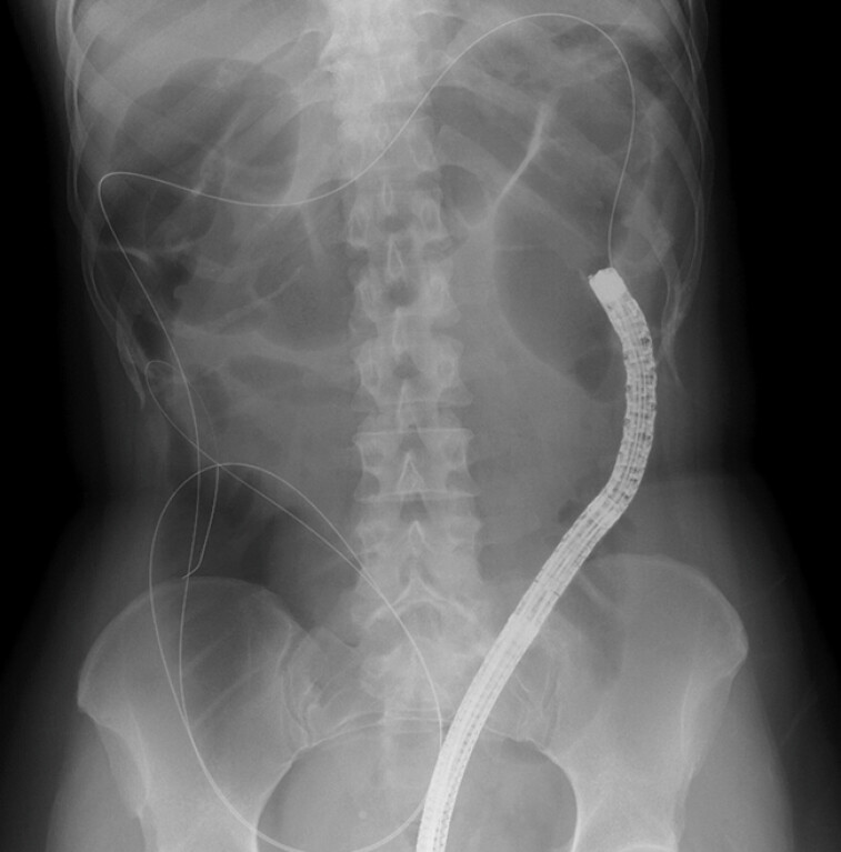 Fig. 2
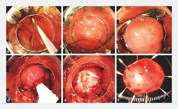 Fig. 3
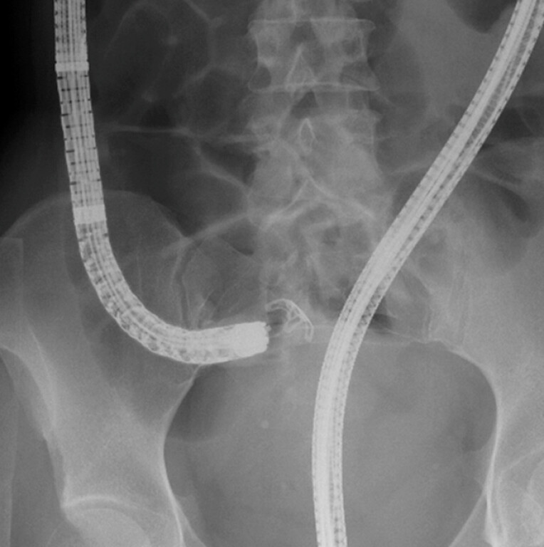 Fig. 4
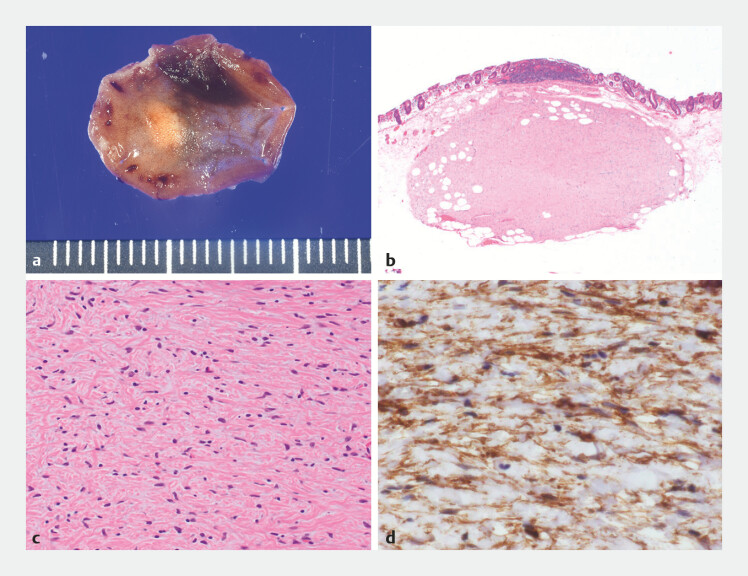 Fig. 5
Peer Reviews
No public reviews on file for this paper yet. If you reviewed it on a platform where reviews are public (OpenReview, ICLR, NeurIPS, ICML), you can paste yours below so the community can read it here.
Videos
No videos yet. Explain this paper in a talk, walkthrough, or lecture? Add one.
Taxonomy
TopicsGastrointestinal Tumor Research and Treatment · Neuroendocrine Tumor Research Advances · Gastrointestinal disorders and treatments
A 48-year-old woman was referred to our hospital for examination and treatment of a cecal submucosal tumor. Colonoscopy was performed for pretreatment screening. The lesion in the cecum measured 5 mm, and the overlying mucosa was intact ( Fig. 1 ). Conventional endoscopic mucosal resection (EMR) or endoscopic submucosal dissection (ESD) would have carried a high risk of perforation and obscured the vertical margin 1 . We therefore decided to perform EMR with over-the-scope (OTS) clips (EMRO).
Colonoscopic images during pretreatment screening showing: a a submucosal tumor measuring 5 mm located in the cecum; b the tumor border revealed on indigo carmine staining.
During the colonoscopy for pretreatment screening, insertion was time-consuming and the patient experienced pain owing to postoperative adhesions, meaning delivery of the OTS clip might have been difficult. We therefore planned to perform guidewire-assisted EMRO (GA-EMRO). First, we inserted the scope into the cecum, and the proximal side of the target lesion was marked using a snare tip. We then placed the guidewire, and carefully withdrew the scope ( Fig. 2 ). We attached an OTS clip to the tip of the scope and safely reinserted it into the cecum with guidewire assistance. The lesion, including the marking, was suctioned into the cap. The OTS clip was successfully deployed, and the lesion was resected with a snare without complications ( Fig. 3 and Fig. 4 ; Video 1 ). Histopathologic examination confirmed a CD34-positive spindle tumor ( Fig. 5 ), with immunohistochemical analysis showing that the spindle cells were positive for CD34 and negative for α-SMA, desmin, c-kit, S-100, and factor XIIIa.
Radiographic image showing the inserted guidewire at the tip of the scope, which was confirmed, on imaging, as remaining in the cecum as the scope was carefully withdrawn.
Images of guidewire-assisted endoscopic mucosal resection with over-the-scope (OTS) clips (GA-EMRO) being performed for a submucosal tumor in the cecum showing: a the scope with the mounted OTS clip being carefully inserted following the guidewire; b the cecal lesion and the previously placed mark; c the lesion and the marking suctioned into the cap; d the successfully deployed OTS clip and the lesion resected with a snare; e the defect post-resection; f the en bloc resected specimen.
Radiographic image showing the deployment of the over-the-scope clip.
Images of the pathologic examination showing: a the macroscopic appearance; b, c on histopathologic appearance after hematoxylin and eosin (H&E) staining: b a clearly demarcated tumor in the submucosal layer (low power magnification); c the presence of spindle cells (high power magnification); d positivity for CD34 on immunohistochemical staining.
Guidewire-assisted endoscopic mucosal resection with over-the scope clips (GA-EMRO) is performed for a submucosal tumor in the cecum.Video 1
A limitation of OTS clips is that they restrict the endoscopic view when attached to the tip of the scope, and delivering the OTS clip may also be problematic 2 3 . EMRO has been reported to be a safe technique for the treatment of duodenal neuroendocrine tumors 4 . We performed GA-EMRO for a submucosal tumor in the cecum. Guidewire-assisted delivery is a valuable and safe method for OTS clip placement in the cecum, and the GA-EMRO technique can facilitate OTS clip treatment of lesions in the proximal large intestine.
Endoscopy_UCTN_Code_TTT_1AQ_2AD_3AF
The reference list from the paper itself. Each links out to its DOI / PubMed record.
- 1Minami S Fukunaga S Mukasa M Novel endoscopic approach for safe and effective resection of duodenal neuroendocrine tumor Endoscopy 202456 E 961E 96210.1055/a-2440-636239515771 PMC 11549000 · doi ↗ · pubmed ↗
- 2Akamine E Asai S Jimbo H Guidewire-assisted method to achieve hemostasis in colonic diverticular bleeding in the ascending colon Endoscopy 202053 E 120E 12110.1055/a-1216-041332707581 · doi ↗ · pubmed ↗
- 3Nishiyama N Mori H Kobara H Safe guidewire-assisted method of over-the-scope clip delivery for the bleeding in the small intestine Endoscopy 201547 E 590E 59110.1055/s-0034-139338226671538 · doi ↗ · pubmed ↗
- 4Tashima T Ryozawa S Tanisaka Y Endoscopic resection using an over-the-scope clip for duodenal neuroendocrine tumors Endoscopy 202109 E 659E 66610.1055/a-1374-6141 PMC 806222833937505 · doi ↗ · pubmed ↗