Underwater endoscopic papillectomy of a duodenal adenoma extending to the papilla using a forward-viewing endoscope
Takashi Yamamoto, Yasushi Yamasaki, Yuki Fujii, Kazuyuki Matsumoto, Motoyuki Otsuka

Abstract
Genes, proteins, chemicals, diseases, species, mutations and cell lines named across the full text — each resolved to its canonical identifier and authoritative record.
Click any figure to enlarge with its caption.
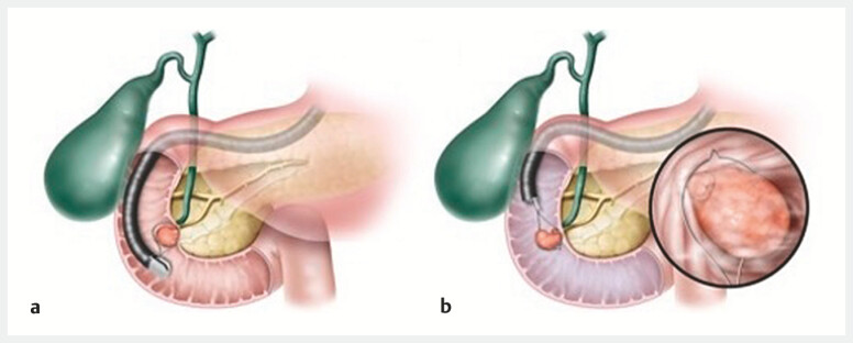 Fig. 1
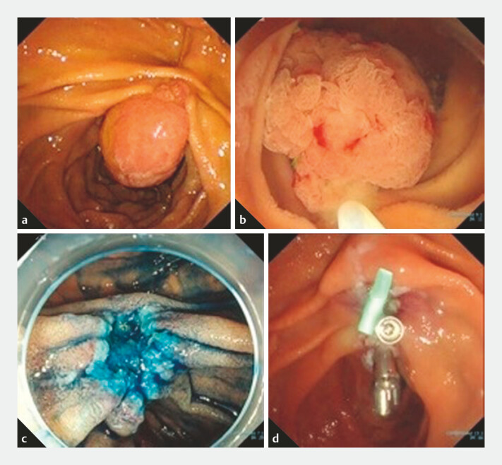 Fig. 2
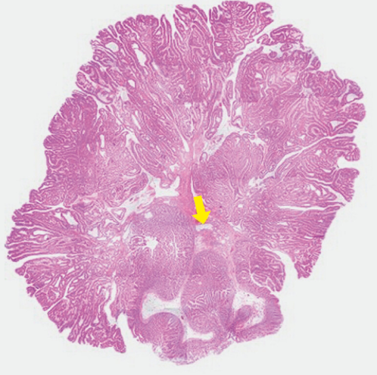 Fig. 3
Peer Reviews
No public reviews on file for this paper yet. If you reviewed it on a platform where reviews are public (OpenReview, ICLR, NeurIPS, ICML), you can paste yours below so the community can read it here.
Videos
No videos yet. Explain this paper in a talk, walkthrough, or lecture? Add one.
Taxonomy
TopicsGallbladder and Bile Duct Disorders · Pancreatic and Hepatic Oncology Research · Esophageal and GI Pathology
Endoscopic resection of duodenal adenomas extending to the papilla is challenging 1 . Endoscopic papillectomy using an oblique-viewing endoscope is generally performed for ampullary adenomas; however, the vertical approach and snaring of the lesion carry a risk of muscle layer involvement, particularly in large lesions (>20 mm) or nonampullary adenomas extending to the papilla 2 . In contrast, a forward-viewing endoscope allows for a horizontal approach, enabling shallower resection and reducing the risk of perforation ( Fig. 1 ). We herein report a successful case of endoscopic papillectomy for a large duodenal adenoma extending to the papilla, performed using a forward-viewing endoscope in combination with the underwater technique ( Video 1 ).
Schematic showing: a vertical snaring with an oblique-viewing endoscope; b horizontal snaring using a forward-viewing endoscope with the underwater technique.
Underwater endoscopic papillectomy is performed using a forward-viewing endoscope for a 25-mm duodenal adenoma extending to the papilla.Video 1
A woman in her 50s was referred to our hospital with a 25-mm duodenal adenoma extending to the papilla ( Fig. 2 a ). The lesion was primarily located on the distal side of the papilla, with extension to the papilla itself, posing a risk of intraoperative perforation if conventional endoscopic papillectomy with an oblique-viewing endoscope were performed. Therefore, we used a forward-viewing endoscope (PCF-290TI; Olympus, Tokyo, Japan). Underwater, the lesion floated owing to buoyancy and low intraduodenal pressure, facilitating easy snaring of the entire lesion with a horizontal approach. The lesion was resected en bloc using electrocautery, without any adverse events occurring ( Fig. 2 b, c ). After clip closure of the distal side of the mucosal defect had been completed, the scope was exchanged for an oblique-viewing endoscope (TJF-Q290V; Olympus), and a pancreatic stent was placed ( Fig. 2 d ). Histological examination confirmed a duodenal adenoma extending to the papilla, with negative horizontal and vertical margins; no tumor invasion was identified ( Fig. 3 ).
Endoscopic images during underwater endoscopic papillectomy using a forward-viewing endoscope showing: a a 25-mm duodenal adenoma involving the papilla of Vater (view with an oblique-viewing endoscope); b the lesion, including the papilla, snared during underwater endoscopic papillectomy (forward-viewing endoscope); c the mucosal defect after underwater endoscopic papillectomy stained with indigo carmine; d appearance after insertion of a pancreatic stent into the main pancreatic duct and closure of the mucosal defect with clips.
Histological examination of the resected lesion showing no tumor invasion into the main pancreatic duct (yellow arrow).
For ampullary adenomas, a vertical approach is crucial for deeper resection because of potential invasion into the bile and pancreatic ducts; however, a horizontal approach with a forward-viewing endoscope underwater may be more appropriate for duodenal adenomas extending to the papilla.
Endoscopy_UCTN_Code_TTT_1AR_2AF
The reference list from the paper itself. Each links out to its DOI / PubMed record.
- 1Itoi T Ryozawa S Katanuma A Clinical practice guidelines for endoscopic papillectomy Dig Endosc 20223439441110.1111/den.1423335000226 · doi ↗ · pubmed ↗
- 2Tonai Y Takeuchi Y Akita H Iatrogenic duodenal perforation during underwater ampullectomy: endoscopic repair using polyglycolic acid sheets Endoscopy 201648 E 97E 9826975296 10.1055/s-0042-103926 · doi ↗ · pubmed ↗