Benign Osseous Metaplasia of the Breast: A Rare Breast Lesion—Case Report With Clinicopathologic Features and Review of the Literature
Mukund Tinguria, Angela Fleming

TL;DR
A rare case of benign osseous metaplasia in the breast is reported, highlighting the importance of histopathological analysis for accurate diagnosis.
Contribution
This case report adds to the limited literature on primary benign osseous metaplasia of the breast without associated breast disease.
Findings
The lesion was confirmed as benign osseous metaplasia through histologic and immunohistochemical analysis.
Immunohistochemistry ruled out metaplastic carcinoma by showing negative staining for epithelial markers.
The case demonstrates the necessity of thorough histopathological evaluation for accurate diagnosis.
Abstract
Benign osseous metaplasia of the breast is an extremely rare breast lesion. This is a report of a 79-year-old woman who presented with a right breast lesion. The lesion was found incidentally on computed tomography (CT) scan examination of the chest. Subsequent mammogram showed coarse calcification within a round circular area measuring approximately 12 mm in size. Ultrasound examination showed an ill-defined 9 × 9 × 9 mm hypoechoic lesion with calcification and internal vascularity. Histologic examination of the excised lesion showed features of benign osseous metaplasia. There was no evidence of atypia and malignancy. The subsequent immunohistochemistry confirmed the diagnosis. The immunohistochemical staining for epithelial markers—pancytokeratin (AE1/AE3), Cam 5.2, HMWCK (34Be12), and CK5—was negative in the stromal component, which ruled out a metaplastic carcinoma. Osseous…
Genes, proteins, chemicals, diseases, species, mutations and cell lines named across the full text — each resolved to its canonical identifier and authoritative record.
Click any figure to enlarge with its caption.
 Figure 1
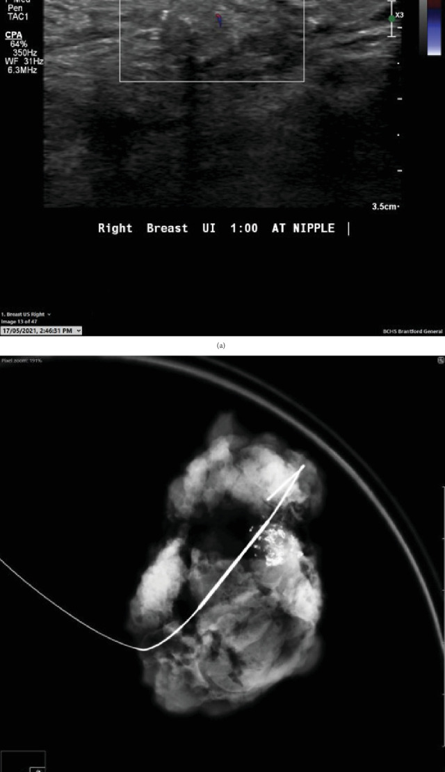 Figure 2
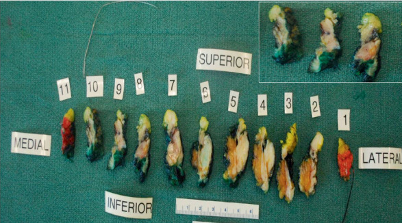 Figure 3
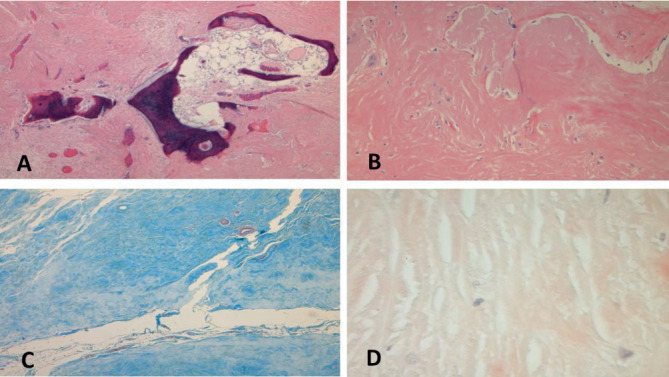 Figure 4
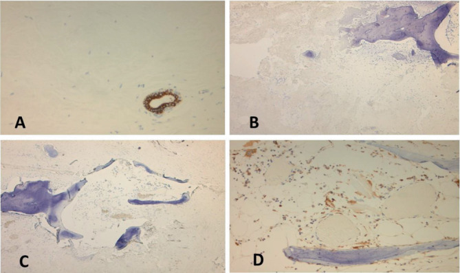 Figure 5
Peer Reviews
No public reviews on file for this paper yet. If you reviewed it on a platform where reviews are public (OpenReview, ICLR, NeurIPS, ICML), you can paste yours below so the community can read it here.
Videos
No videos yet. Explain this paper in a talk, walkthrough, or lecture? Add one.
Taxonomy
TopicsMedical Imaging and Pathology Studies · Breast Lesions and Carcinomas · Heterotopic Ossification and Related Conditions
1. Introduction
Osseous metaplasia, also referred to as metaplastic ossification or heterotopic bone formation, can occur in a variety of neoplastic and non-neoplastic conditions at multiple body sites [1–5]. Osseous metaplasia in the breast is uncommon. The occurrence of bone and cartilage has been described in association with both benign and malignant breast neoplasms. Osseous metaplasia in the absence of breast disease is, however, exceedingly rare, with less than 10 cases being described in the medical literature [6–12]. The entity is diagnostically challenging, as it may mimic a neoplasm both clinically and on imaging studies. The aim of the report is to present clinicopathological features of primary nontumoral osseous metaplasia of the breast, emphasizing the importance of detailed pathological examination to arrive at a correct diagnosis. The report also describes differential diagnoses and pathogenesis theories of this rare entity.
2. Materials and Methods
2.1. Case Report
A 79-year-old woman presented to the hospital with few months' history of weight loss and constipation. The colonoscopy revealed mild to moderate pancolonic diverticulosis and anal stenosis with tight anal sphincter. The esophagogastroduodenoscopy revealed a small hiatus hernia. There was no evidence of ulcer or a mass lesion in the gastrointestinal tract. To further investigate the weight loss, imaging procedure studies were carried out (see Table 1 for patient's diagnostic work-up). The CT scan examination did not show any tumor in the lungs, abdomen, and pelvis. A focus of increased attenuation was noted in the upper half of the right breast (Figure 1) and breast imaging procedure studies were recommended to further investigate this abnormal area. Subsequent breast mammogram showed coarse calcification within a round circular area measuring approximately 12 mm in size in the upper half of the right breast in subareolar region (Figure 1). This area conformed geographically and size-wise to the area of hyperdensity seen on CT scan of thorax. Review of previous imaging study documents showed that this area was present in the CT scan examination carried out 3 years before; however, it was smaller and less calcified that time. The differential diagnoses on mammogram included a calcified cyst. Ultrasound (US) examination of the right breast showed an ill-defined 9 × 9 × 9 mm isoechoic to minimally hypoechoic lesion with internal and peripheral calcifications and internal vascularity (Figure 2). In addition, mild ductal ectasia and a 5-mm cyst were noted at 9 o'clock radian. No other lesions were present. US examination diagnosis was that of a suspicious abnormality (BIRADS 4), and US-guided biopsy of the lesion was recommended for definitive pathologic diagnosis.
Imaging procedure studies of the left breast demonstrated a 1.5 cm nodule in the left axilla, which was present on numerous prior imaging studies. Imaging studies favored the diagnosis of a benign lesion such as a cyst or a lymph node.
2.2. Pathology
The needle core biopsy from the right breast lesion showed dense sclerosis with areas of osseous metaplasia. Mild inflammatory infiltrate consisting of lymphocytes, plasma cells, histiocytes, and foreign body type giant cells was present. Occasional benign normal breast ducts were noted. There was no evidence of atypia or malignancy. As osseous metaplasia can be associated with a wide variety of benign and malignant lesions, US-guided wire localization excision of the lesion was carried out for a definite pathologic diagnosis. The specimen mammogram after wire localization excision demonstrated a partially calcified nodule (Figure 2b).
2.3. Macroscopic Features
The specimen measured 6.2 × 4.7 × 1.8 cm. The area of concern was delineated with a localization wire. The specimen was sliced into 11 slices. Gross examination showed scattered ill-defined areas of fibrosis, but no obvious gross tumor was noted (Figure 3). The area of concern seen on the specimen mammogram did not look different from the surrounding breast parenchyma. As no tumor or an obvious lesion was identified on gross examination, the entire specimen was submitted for histologic examination.
2.4. Microscopic Features
Microscopic examination showed stromal sclerosis with dense paucicellular hyaline stroma and benign osseous metaplasia. Osseous metaplasia was characterized by mature bone fragments set in a fibrous stroma forming a mass-like lesion (Figure 4). Few scattered plasma cells, giant cells, and histiocytes were noted. There was no evidence of atypia and malignancy. The fibrous background was negative for amyloid on Congo red stain, which ruled out amyloid tumor (Figure 4). It showed features of collagen on trichrome staining (Figure 4). Epithelial markers pancytokeratin (AE1/AE3), Cam 5.2, HMWCK (34Be12), and CK5 were negative in the stromal component, which ruled out a metaplastic carcinoma (Figures 5, 5, 5, and 5). The surrounding breast showed features of fibrocystic change. The microscopic diagnosis was that of benign osseous metaplasia. Follow-up of the patient was recommended by regular breast imaging studies considering the rarity of the lesion with limited data available in the literature.
3. Discussion
Osseous metaplasia, also referred to as metaplastic ossification or heterotopic bone formation, can occur in a variety of neoplastic and non-neoplastic conditions at multiple body sites such as the endometrium, ovaries, thyroid, parathyroid, urinary bladder, thymus, scar tissue, and gastrointestinal tract, among others [1–5]. However, osseous metaplasia in the breast is rare. Bonet may have been the first to record a case of osseous metaplasia in the breast, describing a tumor that could not be cut with a knife [13]. Since then, there have been over 200 reports of osseous metaplasia in the breast associated with primary breast cancers, metastatic tumors of the breast, benign breast tumors, a variety of non-neoplastic conditions, and very rarely as an isolated finding.
Benign breast tumors showing focal osseous metaplasia include fibroadenoma, pleomorphic adenoma, benign mesenchymoma, phyllodes tumor, amyloid tumor, fat necrosis, atypical ductal hyperplasia, lipogranuloma, cholesterol granuloma, papilloma, hemangioma, breast abscess, chronic mastitis, fasciitis ossificans, and breast implant [14–30].
Malignant neoplasms showing osseous metaplasia include metaplastic breast carcinoma, invasive ductal carcinoma, fibrosarcoma, osteosarcoma, osteochondrosarcoma, malignant phyllodes tumor, and metastatic osteosarcoma to breast [19, 20, 31–38].
Osseous metaplasia in the breast in the absence of any other breast disease is extremely rare. Review of the literature revealed less than 10 cases of this rare entity [6–12]. The clinicopathologic features of benign osseous metaplasia of the breast are described in the table (Table 2). The age in reported cases ranged from 37 to 71 years. Most patients presented with a breast lump.
The imaging findings in primary osseous metaplasia of the breast are variable. In our case, mammogram demonstrated coarse calcification within a round circular area, and subsequent US showed an ill-defined hypoechoic lesion measuring 9 × 9 × 9 mm with internal and peripheral calcification and internal vascularity. The impression on imaging studies was of a suspicious lesion. Imaging findings in the reported cases of benign osseous metaplasia of the breast include suspicious looking lesions on mammogram and US examination [7], suspicious cluster of calcifications [6], cystic mass with areas of calcification [8], and features suggesting calcified fibroadenoma [9, 10].
Pathologic examination may or may not reveal gross lesions; when present, it is usually a well-circumscribed nodular lesion. Histologic examination shows densely sclerosed stroma with calcification and ossification with bone matrix, with osteocytes, osteoclasts, and spindle cells. Immunohistologic studies show negativity for epithelial markers such as pancytokeratin (AE1/AE3), Cam 5.2, HMWCK, MNF, CK5/6, p63, and CK14, which rule out metaplastic carcinoma [7, 12]. Pathologic examination is crucial for a definite diagnosis, as osseous metaplasia can be seen in a wide range of benign and malignant breast lesions. Benign osseous metaplasia, in the absence of other breast pathology, remains a diagnosis of exclusion when no other associated lesions are found on detailed pathological examination. The value of thorough sampling for pathologic examination cannot be emphasized to rule out foci of malignancy, considering prognostic and therapeutic implications.
Metaplastic breast carcinoma is one of the important differential diagnoses and needs to be ruled out by morphologic and appropriate immunohistologic studies. Metaplastic breast carcinoma represents a heterologous group of breast carcinomas characterized by differentiation of neoplastic epithelium towards squamous cells or mesenchymal appearing elements including but not limited to spindle, chondroid, and osseous cells. When these tumors lack a ductal carcinoma in situ (DCIS) or conventional mammary carcinoma component, a panel of immunohistochemical stains is required to confirm the presence of epithelial differentiation. Metaplastic carcinomas can be epithelial only carcinomas, pure (monophasic) sarcomatoid carcinomas, and biphasic epithelial and sarcomatoid carcinomas. Epithelial only carcinomas include low- and high-grade adenosquamous carcinomas and squamous cell carcinomas. Pure (monophasic) sarcomatoid carcinomas include fibromatosis-like metaplastic carcinoma and spindle cell carcinoma. Heterologous mesenchymal components include chondroid, osseous, rhabdomyosarcomatous, angiosarcomatous, and neurological differentiation singly or in combination. Mesenchymal components can show a wide spectrum of atypia ranging from minimal atypia to frank malignancy. The identification of the epithelial component may require extensive sampling and immunohistochemistry using broad spectrum keratin markers. Immunohistochemistry carried out in our case did not show positivity for epithelial markers pancytokeratin (AE1/AE3), Cam 5.2, CK5, HMWCK (34Be12), and p63, which ruled out metaplastic carcinoma. Smooth muscle myosin heavy chain (SMMHC-1) and p63 staining highlighted myoepithelial cells in benign ducts.
The pathogenesis of osseous metaplasia is yet to be completely understood. Suggested mechanisms include the transformation of stromal fibroblasts into osteoblasts, epithelial–stromal metaplasia, and ossification of calcific debris. The role of stem cells, cytokines, growth factors, extracellular matrix, and bone morphogenic proteins has been postulated [15]. In non-neoplastic lesions, ossification may be related to the underlying inflammatory processes such as osteoma cutis, which is related to chronic folliculitis [39]. Heterotopic bone develops when stromal fibroblasts are transformed by metaplasia into osteoblasts, and this metaplastic bone may arise directly from fibrous tissue or it can be secondary to cartilage formation [12]. It has been postulated that drug, metabolite, hormonal, and genetic factors also play a role in the development of osseous metaplasia [40].
4. Conclusion
Benign nontumoral osseous metaplasia of the breast is an extremely rare entity and may pose a diagnostic challenge clinically and on imaging studies. As clinical and imaging studies cannot differentiate between neoplastic and non-neoplastic lesions as well as benign and malignant neoplasms with certainty, histologic examination is necessary for definitive diagnosis. It is important for the pathologist to rule out other causes of osseous metaplasia in the breast before making a diagnosis of primary benign osseous metaplasia, which essentially remains a diagnosis of exclusion.
The reference list from the paper itself. Each links out to its DOI / PubMed record.
- 1Bahceci M. Demirel L. C. Case Report: Osseous Metaplasia of the Endometrium: A Rare Cause of Infertility and Its Hysteroscopic Management Human Reproduction 199611112537253910.1093/oxfordjournals.humrep.a 0191542-s 2.0-00304437908981150 · doi ↗ · pubmed ↗
- 2Singh P. Kapur K. Singla S. Naz N. Endometrial Osseous Metaplasia and Mature Bone Formation With Extramedullary Hematopoiesis Journal of Human Reproductive Sciences 201141565710.4103/0974-1208.823632-s 2.0-7996026061521772743 PMC 3136072 · doi ↗ · pubmed ↗
- 3Chun S. Hong R. Jung A. Osseous Metaplasia With Mature Bone Formation of the Thyroid Gland: Three Case Reports Oncology Letters 20136497797910.3892/ol.2013.14752-s 2.0-8488159253524137448 PMC 3796393 · doi ↗ · pubmed ↗
- 4Byard R. Thomas M. Osseous Metaplasia Within Tumours. A Review of 11 Cases Annales de Pathologie 19888164663134902 · pubmed ↗
- 5Tica V. Postolache I. Boșoteanu M. Endometrial Osseous Metaplasia — A Rare Cause of Infertility With Unknown Etiology Medicina 20235910 p. 180310.3390/medicina 5910180337893521 PMC 10608607 · doi ↗ · pubmed ↗
- 6Gal-Gombos E. Esserman L. Poniecka A. Osseous Metaplasia of the Breast: Diagnosis With Stereotactic Core Biopsy Breast Journal 200281505210.1046/j.1524-4741.2002.08011.x 2-s 2.0-003616065511856163 · doi ↗ · pubmed ↗
- 7Joshi M. Remoundos D. D. Ahmed F. Rees G. Cunnick G. An Unusual Breast Lump: Osseous Metaplasia Case Reports 20132013 bcr 201200823910.1136/bcr-2012-0082392-s 2.0-84875928801 PMC 360399623386492 · doi ↗ · pubmed ↗
- 8Thushara K. Rupashree S. Babu R. Benign Osseous Metaplasia of Breast – A Rare Case Report Annals of International Medical and Dental Research 202061 PT 01PT 02