The role of cardiac imaging in the post-ISCHEMIA world
Julia Niemierko, Ed Nicol, Jonathan R Weir-McCall

TL;DR
This paper reviews how cardiac imaging is used to diagnose stable chest pain, focusing on insights from the ISCHEMIA trial.
Contribution
It provides a synthesis of the ISCHEMIA trial's impact on the role of anatomical and functional imaging in stable chest pain.
Findings
The ISCHEMIA trial redefined the use of anatomical and functional imaging in stable chest pain.
Non-invasive imaging has gained stronger evidence for diagnosing stable chest pain.
The paper contextualizes the ISCHEMIA trial within broader literature on cardiac imaging.
Abstract
The last decade has seen a significant expansion of the evidence supporting the use of non-invasive imaging in the diagnosis of stable chest pain. The most significant of these was the ISCHEMIA trial which redefined the role of anatomical and functional imaging. The current paper examines this trial in the context of the wider literature, to bring together a review of the role of imaging in the patient with stable chest pain.
Genes, proteins, chemicals, diseases, species, mutations and cell lines named across the full text — each resolved to its canonical identifier and authoritative record.
Click any figure to enlarge with its caption.
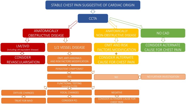 Figure 1
 Figure 2
 Figure 3
 Figure 4
Peer Reviews
No public reviews on file for this paper yet. If you reviewed it on a platform where reviews are public (OpenReview, ICLR, NeurIPS, ICML), you can paste yours below so the community can read it here.
Videos
No videos yet. Explain this paper in a talk, walkthrough, or lecture? Add one.
Taxonomy
TopicsCardiac Imaging and Diagnostics · Advanced MRI Techniques and Applications · Atomic and Subatomic Physics Research