Probing Field Cancerization in the Gastrointestinal Tract Using a Hybrid Raman and Partial Wave Spectroscopy Microscope
Elena Kriukova, Mikhail Mazurenka, Sabrina Marcazzan, Markus Tschurtschenthaler, Gerwin Puppels, Sarah Glasl, Dieter Saur, Moritz Jesinghaus, Marialena Pouliou, Marios Agelopoulos, Apostolos Klinakis, Michael Quante, Jorge Ripoll, Vasilis Ntziachristos, Dimitris Gorpas

TL;DR
This paper introduces a hybrid microscope combining Raman and partial wave spectroscopy to detect early cancerous changes in the gastrointestinal tract of mice.
Contribution
The novel use of a hybrid RS-PWS microscope improves detection of field cancerization in macroscopically normal tissues.
Findings
Raman spectroscopy detects increased amino acid and decreased lipid/carotenoid signals in tumor-adjacent tissues.
Combining Raman and partial wave spectroscopy improves classification accuracy by up to 9% in mouse models.
Transcriptomic analysis confirms a correlation between optical findings and gene expression changes.
Abstract
Field cancerization (FC) refers to spatially distributed premalignant tissue changes that lead to the appearance of local malignancy, and its detection can improve cancer screening. In this work, we employ combined Raman and partial wave spectroscopy (RS-PWS) to detect FC in gastroesophageal (L2-IL1B) and intestinal (Villin-Cre, Apcfl/wt) tumor mouse models. Using a hybrid RS-PWS microscope, we acquire both molecular and morphological information from macroscopically normal tumor-adjacent tissue and investigate the individual and combined performance of each modality. For data analysis, we use partial least-squares discriminant analysis (PLS-DA). In the normal tissue of L2-IL1B mice, we demonstrate a statistically significant increase (p < 0.001) in Raman band intensities associated with free amino acids and a decrease in bands associated with lipids (p < 0.005) and carotenoids (p <…
Genes, proteins, chemicals, diseases, species, mutations and cell lines named across the full text — each resolved to its canonical identifier and authoritative record.
Click any figure to enlarge with its caption.
 Figure 1
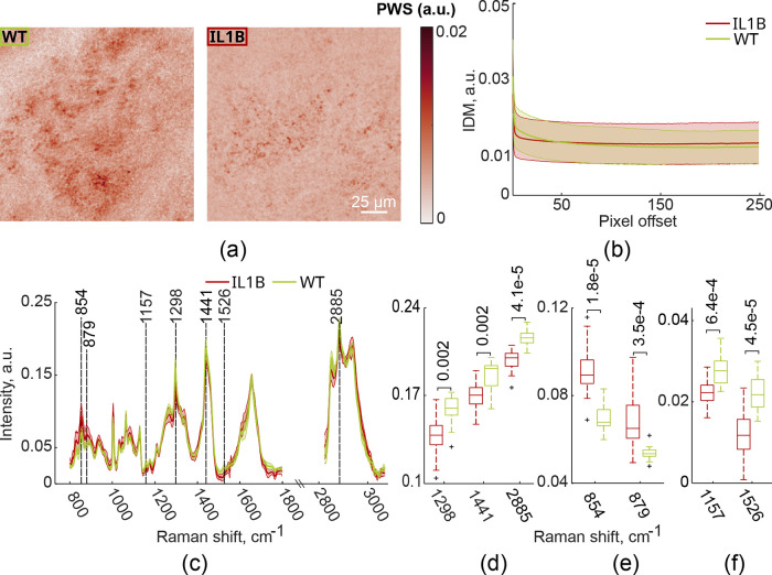 Figure 2
 Figure 3
 Figure 4
 Figure 5
 Figure 6
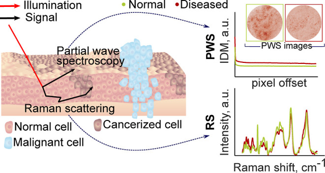 Figure 7
 Figure 8
Peer Reviews
No public reviews on file for this paper yet. If you reviewed it on a platform where reviews are public (OpenReview, ICLR, NeurIPS, ICML), you can paste yours below so the community can read it here.
Videos
No videos yet. Explain this paper in a talk, walkthrough, or lecture? Add one.
Taxonomy
TopicsSpectroscopy Techniques in Biomedical and Chemical Research · Spectroscopy and Chemometric Analyses · Listeria monocytogenes in Food Safety