Membranous Obstruction of the IVC: Role of Advanced Imaging and Intravascular Ultrasound
Rohan Naik, Muhammad Yasir Adeel, Khagendra Dahal, Bruce A. Rheaume, Juyong Lee

TL;DR
This paper presents a case of IVC obstruction caused by membranous blockage and highlights the role of advanced imaging and intravascular ultrasound in diagnosis and treatment.
Contribution
Demonstrates the utility of intravascular ultrasound in diagnosing and managing membranous IVC obstruction.
Findings
A 66-year-old woman had IVC obstruction due to septate membranes.
Membranotomy with balloon angioplasty successfully treated the obstruction.
Intravascular ultrasound was critical for diagnosis and procedural guidance.
Abstract
Inferior vena cava (IVC) obstruction is an underdiagnosed clinical entity. Recognizing obstruction of the IVC can be challenging due to its insidious and often subclinical presentation. A 66-year-old woman presented with chronic bilateral lower extremity swelling. An evaluation revealed septate membranes within the IVC that were causing IVC obstruction. This was successfully treated with membranotomy using balloon angioplasty. We discuss our diagnostic approach and highlight the utility of intravascular ultrasound in making a clinical diagnosis and in guiding management.
Genes, proteins, chemicals, diseases, species, mutations and cell lines named across the full text — each resolved to its canonical identifier and authoritative record.
Click any figure to enlarge with its caption.
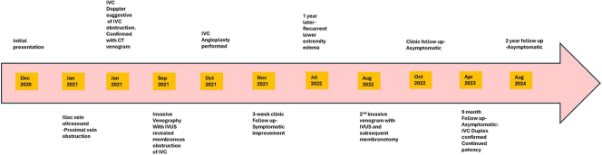 Figure 1
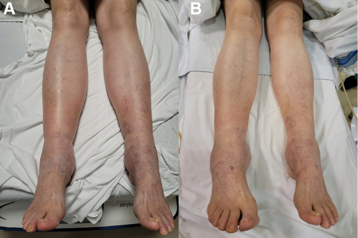 Figure 2
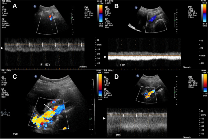 Figure 3
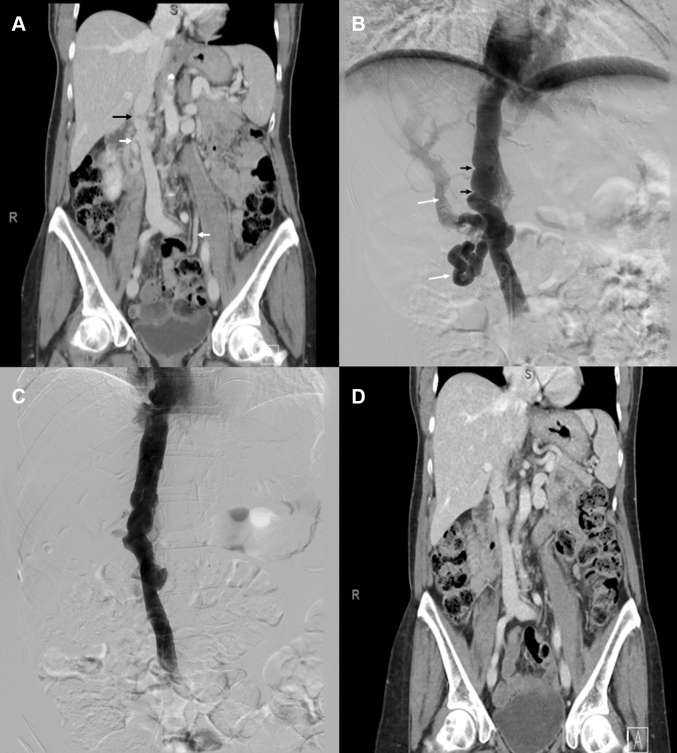 Figure 4
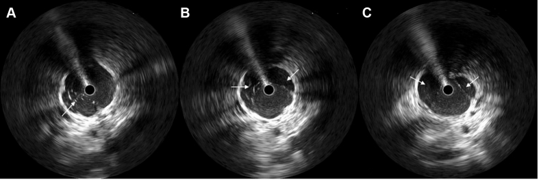 Figure 5
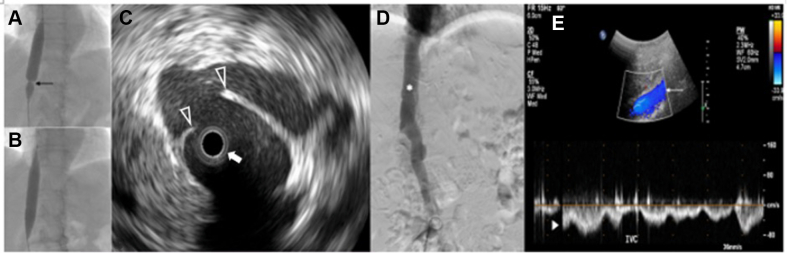 Figure 6
 Figure 7
Peer Reviews
No public reviews on file for this paper yet. If you reviewed it on a platform where reviews are public (OpenReview, ICLR, NeurIPS, ICML), you can paste yours below so the community can read it here.
Videos
No videos yet. Explain this paper in a talk, walkthrough, or lecture? Add one.
Taxonomy
TopicsVenous Thromboembolism Diagnosis and Management · Vascular anomalies and interventions · Central Venous Catheters and Hemodialysis