Radiological Insights into UIP Pattern: A Comparison Between IPF and Non-IPF Patients
Stefano Palmucci, Miriam Adorna, Angelica Rapisarda, Alessandro Libra, Sefora Fischetti, Gianluca Sambataro, Letizia Antonella Mauro, Emanuele David, Pietro Valerio Foti, Claudia Mattina, Corrado Spatola, Carlo Vancheri, Antonio Basile

TL;DR
This study compares radiological features of primary and secondary UIP patterns in lung CT scans to improve diagnostic accuracy.
Contribution
The study identifies distinct radiological markers to differentiate primary UIP/IPF from secondary UIP using expert analysis and AI tools.
Findings
Primary UIP cases showed uniform honeycombing and cranio-caudal fibrosis distribution.
Secondary UIP exhibited patchy fibrosis and irregular GGO distribution, with features like exuberant honeycombing and wedge-shaped fibrosis.
Expert radiologists achieved high sensitivity and specificity in distinguishing primary from secondary UIP.
Abstract
Background/Objectives: This study aims to distinguish radiological differences between primary idiopathic Usual Interstitial Pneumonia (UIP) and secondary UIP patterns Methods: This retrospective study included patients with HRCT findings consistent with a UIP pattern. Final diagnoses were established via multidisciplinary discussion and classified as primary UIP/IPF or secondary UIP, following the 2022 ATS/ERS/JRS/ALAT guidelines. An expert thoracic radiologist (>10 years of experience), blinded to clinical data, reviewed the earliest available HRCT assessing key imaging features: honeycombing (micro-, macro- or exuberant), fibrosis distribution (symmetry, anterior-upper lobe sign, etc.), ground-glass opacities (GGO), dilatation of esophagus. Additionally, AI software AVIEW Build 1.1.46.28-win Coreline (©Coreline Soft Co., Ltd. All Rights Reserved). performed lung texture analysis,…
Genes, proteins, chemicals, diseases, species, mutations and cell lines named across the full text — each resolved to its canonical identifier and authoritative record.
Click any figure to enlarge with its caption.
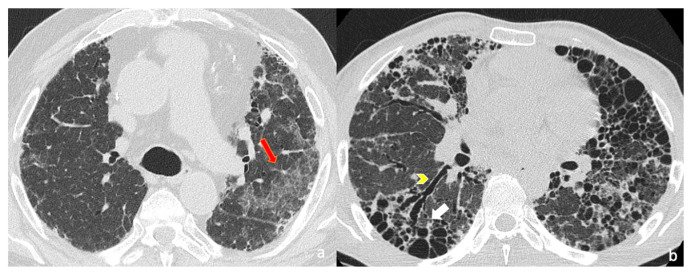 Figure 1
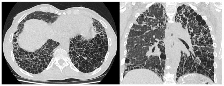 Figure 2
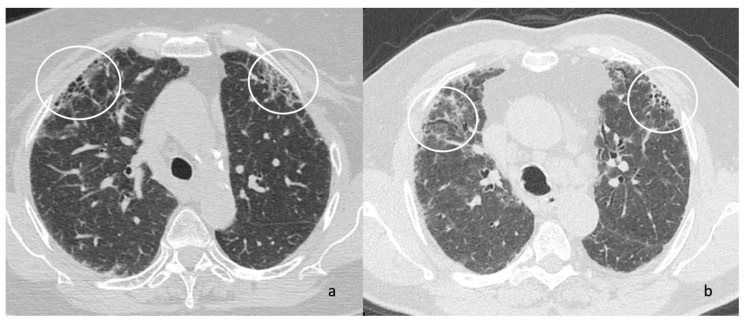 Figure 3
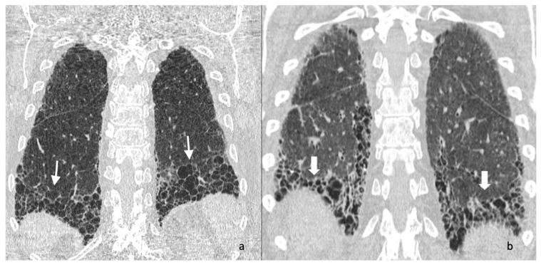 Figure 4
 Figure 5
Peer Reviews
No public reviews on file for this paper yet. If you reviewed it on a platform where reviews are public (OpenReview, ICLR, NeurIPS, ICML), you can paste yours below so the community can read it here.
Videos
No videos yet. Explain this paper in a talk, walkthrough, or lecture? Add one.
Taxonomy
TopicsInterstitial Lung Diseases and Idiopathic Pulmonary Fibrosis · Pneumothorax, Barotrauma, Emphysema · Medical Imaging and Pathology Studies