Opercular Perivascular Space Mimicking a Space-Occupying Brain Lesion: A Short Case Series
Roberts Tumelkans, Cenk Eraslan, Arturs Balodis

TL;DR
This paper presents two cases of a rare brain structure that can be mistaken for a tumor, highlighting the need for accurate diagnosis to avoid unnecessary treatment.
Contribution
The paper contributes two new clinical cases of a recently identified type of perivascular space that mimics brain tumors on imaging.
Findings
The cystic lesions in both patients remained stable over time on follow-up MRI.
The lesions were located near the M2 and M3 segments of the middle cerebral artery in the anterior temporal lobe white matter.
The cases demonstrate the potential for misdiagnosis as low-grade cystic tumors due to their appearance and surrounding edema.
Abstract
A newly recognized fourth type of perivascular space has recently been described in the radiological literature. Despite its growing relevance, many radiologists are still unfamiliar with its imaging characteristics, often leading to misinterpretation as cystic neoplasms. Due to its potential for diagnostic confusion, further studies are necessary—particularly those incorporating high-quality imaging examples across various presentations—to facilitate accurate recognition and classification. Perivascular spaces (PVSs) of the brain are cystic, fluid-filled structures formed by the pia mater and located alongside cerebral blood vessels, particularly penetrating arterioles, venules, and capillaries. Under normal conditions, these spaces are small (typically <2 mm in diameter), but in rare instances, they may become markedly enlarged (>15 mm), exerting a mass effect on adjacent brain…
Genes, proteins, chemicals, diseases, species, mutations and cell lines named across the full text — each resolved to its canonical identifier and authoritative record.
Click any figure to enlarge with its caption.
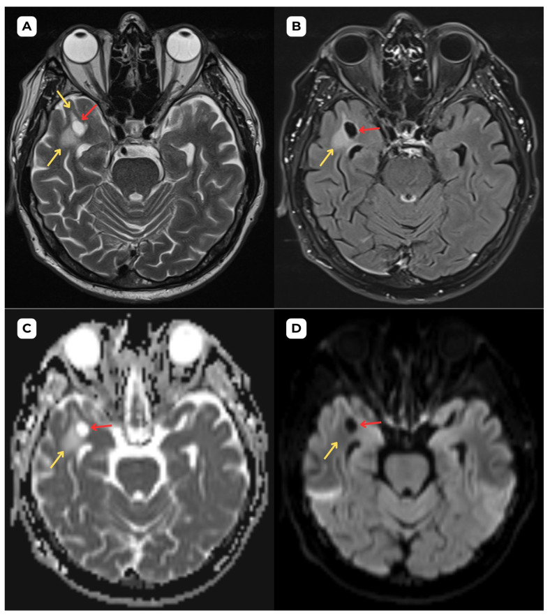 Figure 1
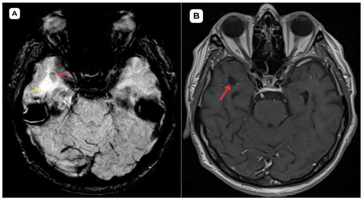 Figure 2
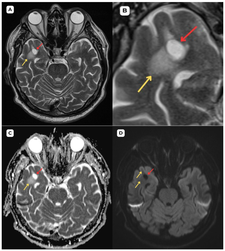 Figure 3
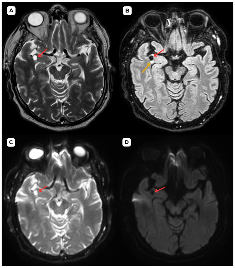 Figure 4
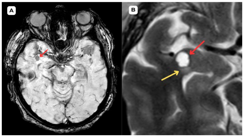 Figure 5
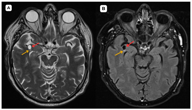 Figure 6
Peer Reviews
No public reviews on file for this paper yet. If you reviewed it on a platform where reviews are public (OpenReview, ICLR, NeurIPS, ICML), you can paste yours below so the community can read it here.
Videos
No videos yet. Explain this paper in a talk, walkthrough, or lecture? Add one.
Taxonomy
TopicsCerebrospinal fluid and hydrocephalus · Spinal Dysraphism and Malformations · Head and Neck Surgical Oncology
The reference list from the paper itself. Each links out to its DOI / PubMed record.
- 1Mc Ardle D.J.T. Lovell T.J.H. Lekgabe E. Gaillard F. Opercular perivascular cysts: A proposed new subtype of dilated perivascular spaces Eur. J. Radiol.202012410883810.1016/j.ejrad.2020.10883831972365 · doi ↗ · pubmed ↗
- 2Capasso R. Negro A. Cirillo S. Iovine S. Puoti G. Cirillo M. Conforti R. Large anterior temporal Virchow–Robin spaces: Evaluating MRI features over the years—Our experience and literature review Clin. Transl. Neurosci.202042514183 X 209052510.1177/2514183 X 20905252 · doi ↗
- 3Doubal F.N. Mac Lullich A.M.J. Ferguson K.J. Dennis M.S. Wardlaw J.M. Enlarged Perivascular Spaces on MRI are a Feature of Cerebral Small Vessel Disease Stroke 20104145045410.1161/STROKEAHA.109.56491420056930 · doi ↗ · pubmed ↗
- 4Wardlaw J.M. Smith C. Dichgans M. Small vessel disease: Mechanisms and clinical implications Lancet Neurol.20191868469610.1016/S 1474-4422(19)30079-131097385 · doi ↗ · pubmed ↗
- 5Lim A.T. Chandra R.V. Trost N.M. Mc Kelvie P.A. Stuckey S.L. Large anterior temporal Virchow-Robin spaces: Unique MR imaging features Neuroradiology 20155749149910.1007/s 00234-015-1491-y 25614333 · doi ↗ · pubmed ↗
- 6Page I. Mc Ardle D.J.T. Gaillard F. Opercular Perivascular Cyst: Old Entity, New Location Neurographics 20211118618810.3174/ng.2000056 · doi ↗
- 7Yu L. Hu X. Li H. Zhao Y. Perivascular Spaces, Glymphatic System and MR Front. Neurol.20221384493810.3389/fneur.2022.84493835592469 PMC 9110928 · doi ↗ · pubmed ↗
- 8Conforti R. Capasso R. Franco D. Russo C. Rinaldi F.O. Pezzullo G. Coluccino S. Brunese M.C. Caiazzo C. Caranci F. Giant Tumefactive Perivascular Space: Advanced Fusion MR Imaging and Tractography Study—A Case Report and a Systematic Review Diagnostics 202313160210.3390/diagnostics 1309160237174993 PMC 10177987 · doi ↗ · pubmed ↗