Biomechanical Analysis and Clinical Study of Augmented Versus Conventional Endoscopic Orbital Decompression for Dysthyroid Optic Neuropathy
Pengsen Wu, Yiheng Wu, Jing Rao, Shenglan Yang, Hongyi Yao, Qingjiang Liu, Yuqing Wu, Shengli Mi, Guiqin Liu

TL;DR
This study uses biomechanical modeling and clinical data to show that augmented orbital decompression surgery is more effective than conventional methods for treating dysthyroid optic neuropathy.
Contribution
A validated finite element analysis model is developed to compare biomechanical outcomes of two orbital decompression techniques.
Findings
Augmented decompression reduces optic nerve stress more effectively than conventional decompression.
Patients in the augmented group showed faster and greater improvements in visual acuity and visual field.
Stress on the optic nerve, eyeball, and orbital wall is higher in dysthyroid optic neuropathy patients than in healthy individuals.
Abstract
Dysthyroid optic neuropathy (DON) represents a severe ocular complication in thyroid eye disease (TED) that can lead to vision loss. Although surgical decompression is a well-established treatment modality, the optimal decompression area remains controversial in orbital decompression surgery. Purpose: This study aims to develop and validate a finite element analysis (FEA) model of DON to compare the biomechanical behavior between patients undergoing conventional or augmented orbital decompression surgery, with potential clinical implications for surgical planning. Methods: FEA models were established using magnetic resonance imaging data from patients with myopathic TED. Pre-disease, preoperative, and postoperative FEA models were developed for both the conventional orbital decompression group and the augmented group, in which the posteromedial floor and the orbital process of the…
Genes, proteins, chemicals, diseases, species, mutations and cell lines named across the full text — each resolved to its canonical identifier and authoritative record.
Click any figure to enlarge with its caption.
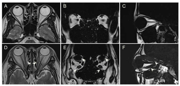 Figure 1
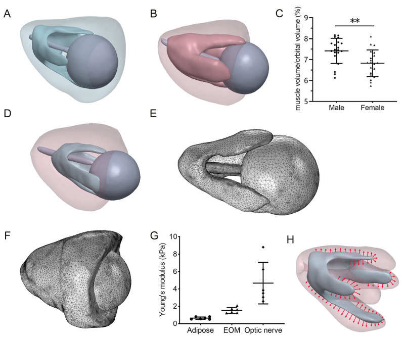 Figure 2
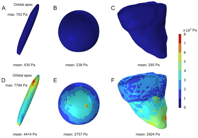 Figure 3
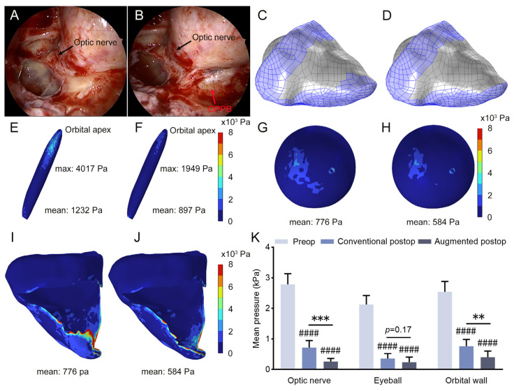 Figure 4
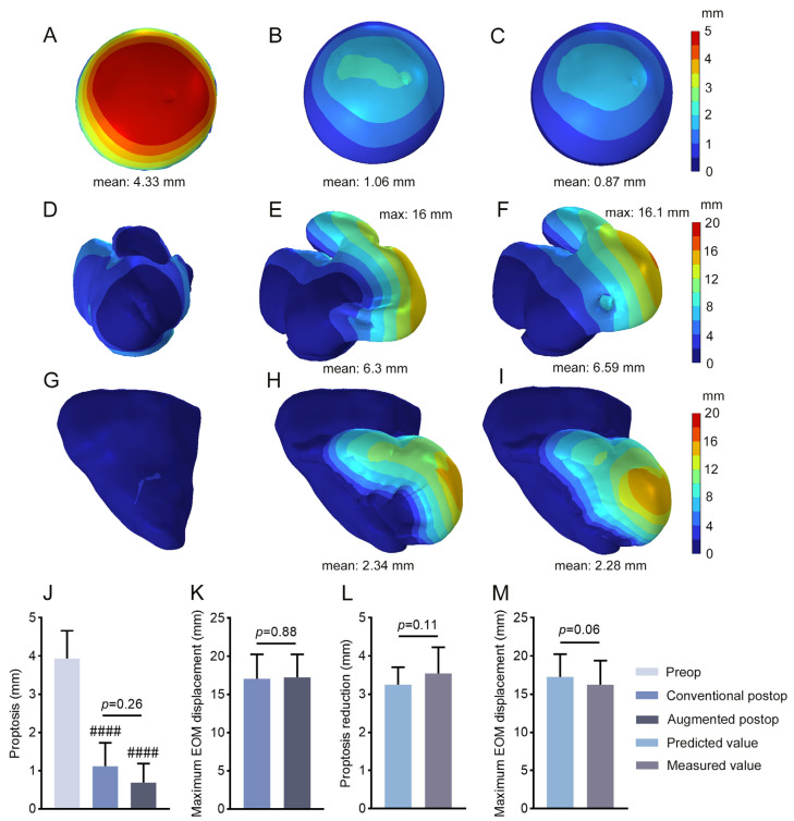 Figure 5
 Figure 6
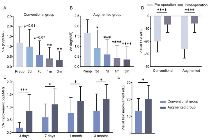 Figure 7
Peer Reviews
No public reviews on file for this paper yet. If you reviewed it on a platform where reviews are public (OpenReview, ICLR, NeurIPS, ICML), you can paste yours below so the community can read it here.
Videos
No videos yet. Explain this paper in a talk, walkthrough, or lecture? Add one.
Taxonomy
TopicsOphthalmology and Eye Disorders · Pituitary Gland Disorders and Treatments · Cerebral Venous Sinus Thrombosis