Adding layers: Environmental and internal modulation of regional skin barrier functions
Joachim W. Fluhr, Razvigor Darlenski

Abstract
Genes, proteins, chemicals, diseases, species, mutations and cell lines named across the full text — each resolved to its canonical identifier and authoritative record.
Click any figure to enlarge with its caption.
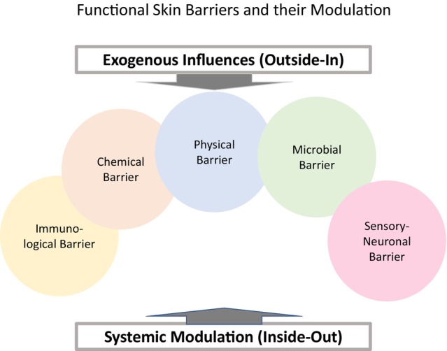 Figure 1
Peer Reviews
No public reviews on file for this paper yet. If you reviewed it on a platform where reviews are public (OpenReview, ICLR, NeurIPS, ICML), you can paste yours below so the community can read it here.
Videos
No videos yet. Explain this paper in a talk, walkthrough, or lecture? Add one.
Taxonomy
TopicsAdvancements in Transdermal Drug Delivery · Dermatology and Skin Diseases · Skin Protection and Aging
The review by Dajnoki et al.1 offers a comprehensive synthesis of current insights into the topographical variability of skin barrier functions. The authors emphasize the complex interplay among chemical, microbial, physical and immunological barriers and explain how these interactions contribute to region‐specific skin vulnerability and disease patterns. To complement and extend the discussion, we propose five key dimensions that could be further integrated into the concept of skin barrier variability.
Environmental conditions such as UV radiation, temperature, humidity, ozone, fine dust and airborne allergens (e.g. pollen) affect skin barrier integrity.2 Recent studies confirm that pollution, particularly ozone and pollen exposure, can induce barrier impairment.3 These factors vary by region, climate and season and directly interact with the skin's surface.
Lifestyle factors—such as engagement in sports, occupational exposure, cultural practices in hygiene and clothing and repetitive mechanical friction—further modulate the barrier. Psychological stress has also been shown to disrupt barrier homeostasis via neuroendocrine pathways.4
The neuronal (sensory) barrier should be considered as active player in barrier function. Sensory nerve fibres influence not only perception but also barrier function, regeneration, inflammation and immune response. Their role is especially relevant in conditions such as sensitive skin and atopic dermatitis.5
The ‘inside‐out’ modulation emphasizes the importance of internal factors in maintaining barrier homeostasis.6 Systemic hydration, nutrition, metabolism and hormonal regulation shape skin reactions to external exposures. Understanding skin function as part of a whole‐body network opens new perspectives for clinical intervention.
Anatomical regions differ functionally and in terms of microenvironmental conditions.7 Corneodesmosomes, vital for the cohesion of the upper stratum corneum, vary in stability and distribution. Occluded areas such as the axillae, groin, anogenital region, submammary folds and plantar surfaces experience elevated moisture, reduced ventilation and altered pH.8 These conditions affect microbiome composition and predispose individuals to infections and maceration.
Hormonal regulation, especially by sex hormones, affects sebaceous gland activity and modifies barrier characteristics in specific regions. Fluctuations during puberty, menstrual cycles and endocrine disorders all contribute to localized barrier behaviour.
In parallel, bacterial adhesion and microbiome diversity vary by site, influencing the establishment and function of local microbial communities. These interactions are key to understanding region‐specific disease patterns.
Building on the foundation laid by Dajnoki et al.,1 incorporating environmental, neuronal, systemic, lifestyle and regional factors will enrich our understanding of skin barrier dynamics and support the development of targeted site‐adapted dermatological strategies (Figure 1).
CONFLICT OF INTEREST STATEMENT
JWF and RD have no conflict of interest to declare related to this article.
The reference list from the paper itself. Each links out to its DOI / PubMed record.
- 1Dajnoki Z , Kapitany A , Eyerich K , Eyerich S , Torocsik D , Szegedi A . Topographical variations in the skin barrier and their role in disease pathogenesis. J Eur Acad Dermatol Venereol. 2025;39(7):1228–1238.10.1111/jdv.20463 PMC 1218850139607016 · doi ↗ · pubmed ↗
- 2Fluhr JW , Darlenski R , Angelova‐Fischer I , Tsankov N , Basketter D . Skin irritation and sensitization: mechanisms and new approaches for risk assessment. 1. Skin irritation. Skin Pharmacol Physiol. 2008;21:124–135.18523410 10.1159/000131077 · doi ↗ · pubmed ↗
- 3Fluhr JW , Stevanovic K , Joshi P , Bergmann KC , Herzog LS , Alwaheed Y , et al. Skin physiology, mucosal functions, and symptoms are modulated by grass pollen and ozone double exposure in allergic patients. Skin Pharmacol Physiol. 2023;36:195–204.36927995 10.1159/000530115 · doi ↗ · pubmed ↗
- 4Choi EH , Brown BE , Crumrine D , Chang S , Man MQ , Elias PM , et al. Mechanisms by which psychologic stress alters cutaneous permeability barrier homeostasis and stratum corneum integrity. J Invest Dermatol. 2005;124:587–595.15737200 10.1111/j.0022-202X.2005.23589.x · doi ↗ · pubmed ↗
- 5Misery L , Bataille A , Talagas M , Le Gall‐Ianotto C , Fouchard M , Huet F , et al. Sensitive skin syndrome: a low‐noise small‐fiber neuropathy related to environmental factors? Front Pain Res (Lausanne). 2022;3:853491.35399156 10.3389/fpain.2022.853491 PMC 8990967 · doi ↗ · pubmed ↗
- 6Elias PM . The skin barrier as an innate immune element. Semin Immunopathol. 2007;29(1):3–14. 10.1007/s 00281-007-0060-9 17621950 · doi ↗ · pubmed ↗
- 7Kleesz P , Darlenski R , Fluhr JW . Full‐body skin mapping for six biophysical parameters: baseline values at 16 anatomical sites in 125 human subjects. Skin Pharmacol Physiol. 2012;25:25–33.21912200 10.1159/000330721 · doi ↗ · pubmed ↗
- 8Zhai H , Maibach HI . Occlusion vs. skin barrier function. Skin Res Technol. 2002;8:1–6.12005114 10.1046/j.0909-752x.2001.10311.x · doi ↗ · pubmed ↗