Effective imaging examination evaluation method for surgical pathological complete responds of head and neck squamous cell carcinoma after neoadjuvant immunochemotherapy
Yudong Ning, Yixuan Song, Yuqin He, Han Li, Yang Liu, Shaoyan Liu

TL;DR
This study finds that using the ROI average value ratio in imaging exams better predicts complete response in head and neck cancer patients after immunochemotherapy compared to standard methods.
Contribution
The study introduces the ROI average value ratio as a more accurate method for evaluating PCR in HNSCC after NIC.
Findings
RECIST v1.1 showed significant discrepancies between imaging and pathology results for PCR in HNSCC patients.
An ROI average value ratio ≥ 1.18 on enhanced CT and ≥ 1.06 on T2-weighted MR imaging was strongly associated with PCR.
The ROI average value ratio demonstrated better diagnostic efficacy for PCR than traditional imaging criteria.
Abstract
To explore an effective method for imaging examinations to evaluate the surgical pathological complete response (PCR) in patients with head and neck squamous cell carcinoma (HNSCC) following neoadjuvant immunochemotherapy (NIC). HNSCC patients who underwent NIC and subsequent surgery from May 2021 to November 2024 were retrospectively analyzed. All patients underwent imaging examination evaluations, including enhanced computed tomography (CT) and enhanced magnetic resonance (MR) imaging both before and after NIC. The average value of the region of interest (ROI) was extracted from the imaging examinations. Clinical parameter-related data were collected. The paired chi-square test was performed to analyze the differences in complete response (CR) between imaging examinations and pathology according to the response evaluation criteria in solid Tumors version 1.1 (RECISTv1.1). The optimal…
Genes, proteins, chemicals, diseases, species, mutations and cell lines named across the full text — each resolved to its canonical identifier and authoritative record.
Click any figure to enlarge with its caption.
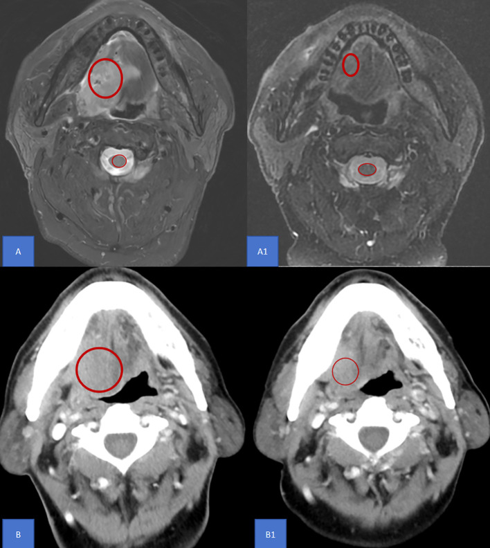 Figure 1
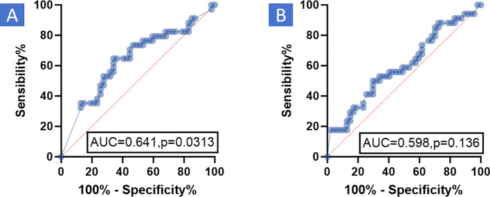 Figure 2
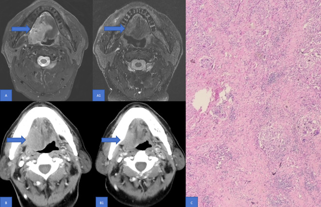 Figure 3
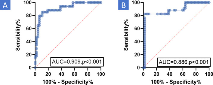 Figure 4
Peer Reviews
No public reviews on file for this paper yet. If you reviewed it on a platform where reviews are public (OpenReview, ICLR, NeurIPS, ICML), you can paste yours below so the community can read it here.
Videos
No videos yet. Explain this paper in a talk, walkthrough, or lecture? Add one.
Taxonomy
TopicsRadiomics and Machine Learning in Medical Imaging · Head and Neck Cancer Studies · Salivary Gland Tumors Diagnosis and Treatment