Chessboard stroke attributed to intracranial atherosclerosis
Raul Medina-Rioja, Juan Carlos Lopez-Hernandez, Enrique Piña-Rosales, Brenda Dzul-García, Andres Mercado-Pompa

Abstract
Genes, proteins, chemicals, diseases, species, mutations and cell lines named across the full text — each resolved to its canonical identifier and authoritative record.
Click any figure to enlarge with its caption.
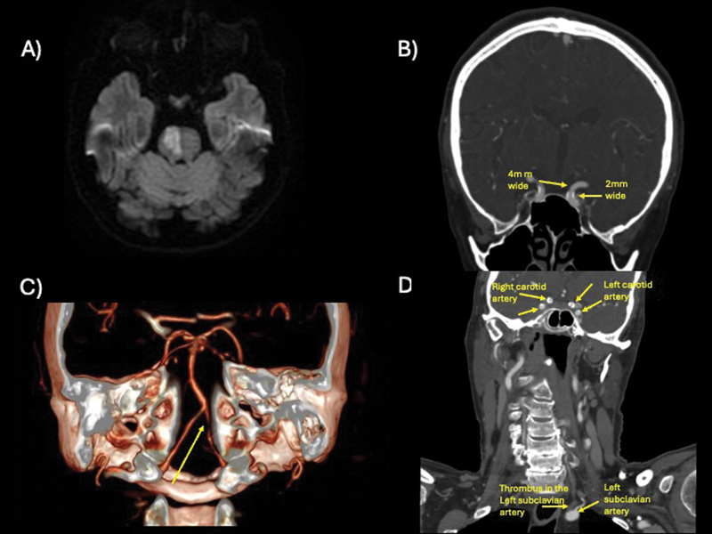 Figure 1
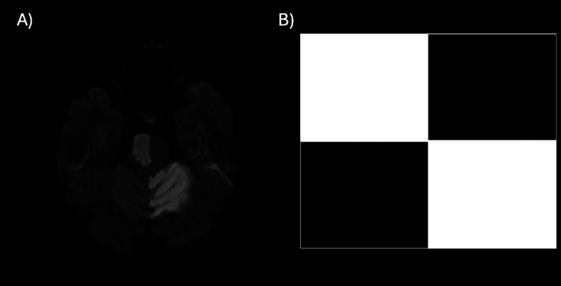 Figure 2
Peer Reviews
No public reviews on file for this paper yet. If you reviewed it on a platform where reviews are public (OpenReview, ICLR, NeurIPS, ICML), you can paste yours below so the community can read it here.
Videos
No videos yet. Explain this paper in a talk, walkthrough, or lecture? Add one.
Taxonomy
TopicsAcute Ischemic Stroke Management · Cerebrovascular and Carotid Artery Diseases · Venous Thromboembolism Diagnosis and Management
A 73-year-old patient presented with sudden double vision and left-sided hemiparesis. A physical exam revealed palsy of the right VI cranial nerve and left arm/leg paresis. A magnetic resonance imaging (MRI) scan showed an acute stroke in the right pons ( Figure 1A ), with the initial workup suggesting intracranial atherosclerosis ( Figure 1B–D ). 1 Other causes were ruled out. The patient was treated with aspirin and clopidogrel and then discharged. He returned the day after with dysarthria and gait instability. A follow-up MRI scan revealed a left cerebellar stroke, creating a chessboard pattern ( Figure 2 ).
( A ) Axial brain magnetic resonance imaging (MRI) scan in diffusion-weighted imaging (DWI) sequence showing an acute stroke located at the right pons. ( B ) Coronal angiotomography showing intracranial atherosclerosis in the internal carotid artery, causing 50% of narrowing of the lumen (4 mm versus 2 mm). ( C ) Tridimensional reconstruction of neck angiotomography showing left vertebral stenosis at the foraminal segment. ( D ) Coronal angiotomography highlighting extra- and intracranial atherosclerosis with a thrombus present in the left subclavian artery.
( A ) Axial brain MRI in DWI sequence showing two strokes located at the right pons and ( B ) the left cerebellum mimicking a chessboard.
The reference list from the paper itself. Each links out to its DOI / PubMed record.