The hidden burden of lysosomal dysfunction: visual decline and microphthalmia in Hunter syndrome
Mateen Sheikh, Ibrahim Thein, Kevin J. Abrams, Leonardo Furtado Freitas

Abstract
Genes, proteins, chemicals, diseases, species, mutations and cell lines named across the full text — each resolved to its canonical identifier and authoritative record.
Click any figure to enlarge with its caption.
 Figure 1
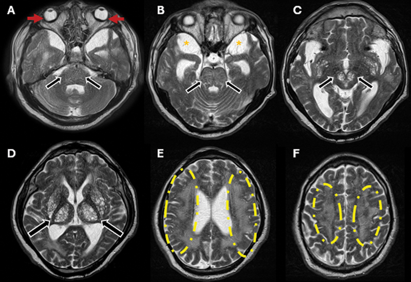 Figure 2
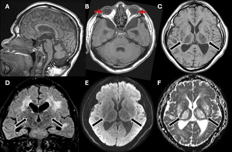 Figure 3
Peer Reviews
No public reviews on file for this paper yet. If you reviewed it on a platform where reviews are public (OpenReview, ICLR, NeurIPS, ICML), you can paste yours below so the community can read it here.
Videos
No videos yet. Explain this paper in a talk, walkthrough, or lecture? Add one.
Taxonomy
TopicsLysosomal Storage Disorders Research · Biomedical Research and Pathophysiology · Cytomegalovirus and herpesvirus research
A 43-year-old male patient with Hunter syndrome (also known as mucopolysaccharidosis type II , MPS II) presented with progressive visual impairment and bilateral upper limb weakness. Multimodal imaging revealed posterior scleral thickening, optic disc edema, and photoreceptor loss, indicating a progressive ophthalmopathy associated with lysosomal storage dysfunction.
While glycosaminoglycan accumulation in the ocular structures has been rarely reported, 1 2 3 the present is the first report of its progression to chronic ophthalmopathy with microphthalmia. Neuroimaging ( Figures 1 2 3 ) demonstrated cervical spinal stenosis with potential dynamic myelopathy, likely contributing to limb weakness. 4 5
Computed tomography (CT) scan of the head in bone window ( A ) and soft tissue window ( B–F ). Intracranial stigmata of mucopolysaccharidosis with enlarged and partially-empty sella turcica (blue arrows), circumferential thickening of the bilateral sclera and microphthalmia (red arrows), prominent cerebrospinal fluid (CSF) in the middle cranial fossae and mega cisterna magna (orange asterisks), ventriculomegaly, and diffuse white matter abnormalities (yellow dashed circles) with multiple prominent perivascular spaces (PVSs; black arrows), predominantly in the posterior subinsular region, putamina, and thalami. Additionally, there is mild diffuse brain atrophy.
Brain magnetic resonance imaging (MRI) scan, axial T2-weighted images ( A–F ). Increased conspicuity of intracranial stigmata of mucopolysaccharidosis. Notable findings include circumferential scleral thickening and microphthalmia (red arrows), attributed to hypointense material representing glycosaminoglycan accumulation. There was also prominent CSF spaces in the middle cranial fossae (orange asterisks), ventriculomegaly, and diffuse white matter abnormalities (yellow dashed circles) with multiple PVSs (black arrows), predominantly in the posterior subinsular region, putamina, and thalami, extending into the upper midbrain. This microcystic appearance is known as the honeycomb pattern.
Brain MRI scan, including sagittal T1-weighted ( A ), axial T1-weighted ( B–C ), coronal fluid-attenuated inversion recovery (FLAIR) ( D ), axial diffusion-weighted imaging ( E ), and apparent diffusion coefficient (ADC) map ( F ). Redemonstration of circumferential scleral thickening and microphthalmia (red arrows). Multiple PVSs (black arrows) with a honeycomb pattern exhibiting signal suppression on T1- and FLAIR-weighted images, along with facilitated diffusion. White matter abnormalities show FLAIR hyperintensity, suggesting gliosis and/or demyelination secondary to lysosomal storage dysfunction and neuronal impairment.
The case herein reported highlights the need for early ophthalmologic and neuroradiologic surveillance to prevent irreversible visual and neurological deterioration in Hunter syndrome.
The reference list from the paper itself. Each links out to its DOI / PubMed record.
- 1Majmudar I P Ismail H O Dang S Gill M K Posterior segment findings in Hunter Syndrome: Case report and review Am J Ophthalmol Case Rep 20243610218910.1016/j.ajoc.2024.10218939498144 10.1016/j.ajoc.2024.102189 PMC 11532303 · doi ↗ · pubmed ↗
- 2Seay M D Lau H Galetta S L Teaching Neuro Images: Scleral thickening and optic disc edema from glycosaminoglycan deposition in Hunter syndrome Neurology 20199213 e 1532 e 153310.1212/WNL.000000000000718330910949 10.1212/WNL.0000000000007183 · doi ↗ · pubmed ↗
- 3Ashworth J L Biswas S Wraith E Lloyd I C The ocular features of the mucopolysaccharidoses Eye (Lond)2006200555356310.1038/sj.eye.670192115905869 10.1038/sj.eye.6701921 · doi ↗ · pubmed ↗
- 4Zafeiriou D I Batzios S P Brain and spinal MR imaging findings in mucopolysaccharidoses: a review AJNR Am J Neuroradiol 2013340151310.3174/ajnr.A 283222790241 10.3174/ajnr.A 2832 PMC 7966323 · doi ↗ · pubmed ↗
- 5Manara R Priante E Grimaldi M Santoro L Astarita L Barone R Brain and spine MRI features of Hunter disease: frequency, natural evolution and response to therapy J Inherit Metab Dis 2011340376378010.1007/s 10545-011-9317-521465231 10.1007/s 10545-011-9317-5 · doi ↗ · pubmed ↗