Epidermoid Cyst of the Cecum Treated by Laparoscopic Colectomy: A Case Report With Histopathology and Literature Review
Ada Firrincieli, Eleonora Nardi, Lavinia Pugliese, Chiara Marconcini, Giovanni Alemanno, Luca Messerini

TL;DR
A rare case of a benign epidermoid cyst in the cecum was successfully treated with laparoscopic surgery and confirmed through histopathology.
Contribution
This case report adds to the limited literature on cecal epidermoid cysts and highlights the importance of histopathology for accurate diagnosis.
Findings
The cyst was confirmed as a benign epidermoid cyst lined by keratinized squamous epithelium.
Imaging techniques failed to provide a conclusive diagnosis, emphasizing the need for histopathological evaluation.
The patient had no malignant findings and recovered well after laparoscopic hemicolectomy.
Abstract
Introduction: Cecal epidermoid cyst (CEC) is a rare and benign lesion; the origin can be acquired or congenital, but the pathogenesis remains unclear. We present a case report of a patient with a cecal cyst treated by hemicolectomy. Histopathology revealed an epidermoid cyst (EC) of the cecum. Case Presentation: A 28-year-old woman was admitted to the hospital with abdominal pain, without significant past medical history. CT and MRI scans were performed, and a large cystic mass in the anterior portion of the pelvic region was detected. Imaging techniques managed to localize the site and dimensions of the neoplasm; however, they did not provide a conclusive diagnosis. The differential diagnosis was made with appendiceal mucocele, duplication cyst, or endometriotic cyst formation. Laparoscopic right hemicolectomy was performed; the mass did not present with any adhesions with the…
Genes, proteins, chemicals, diseases, species, mutations and cell lines named across the full text — each resolved to its canonical identifier and authoritative record.
Click any figure to enlarge with its caption.
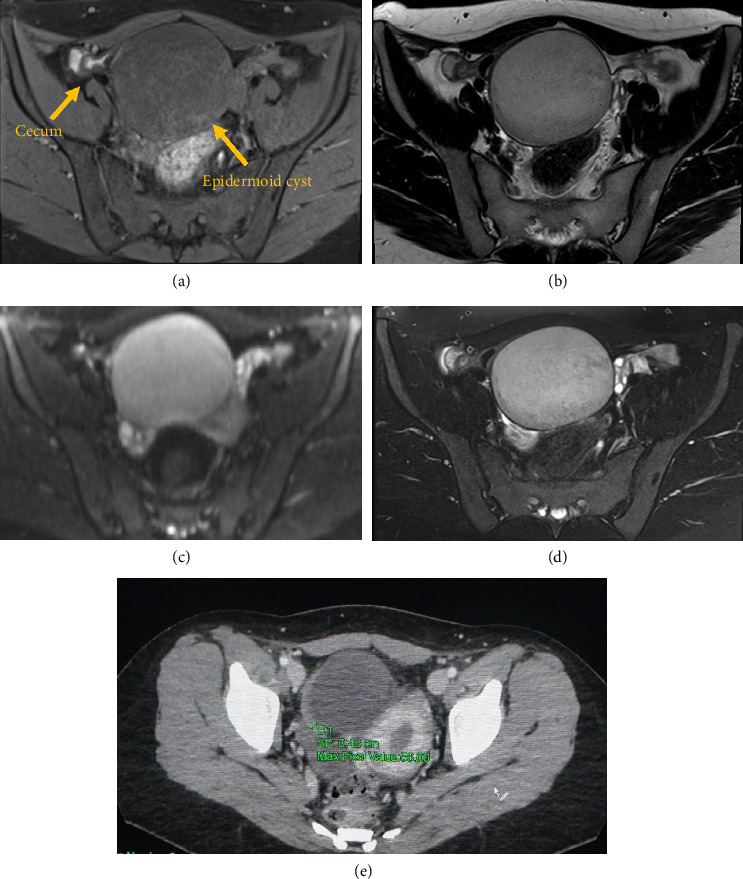 Figure 1
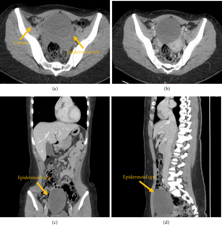 Figure 2
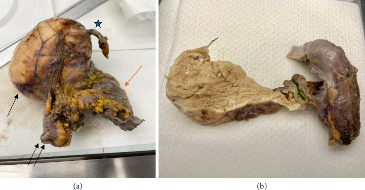 Figure 3
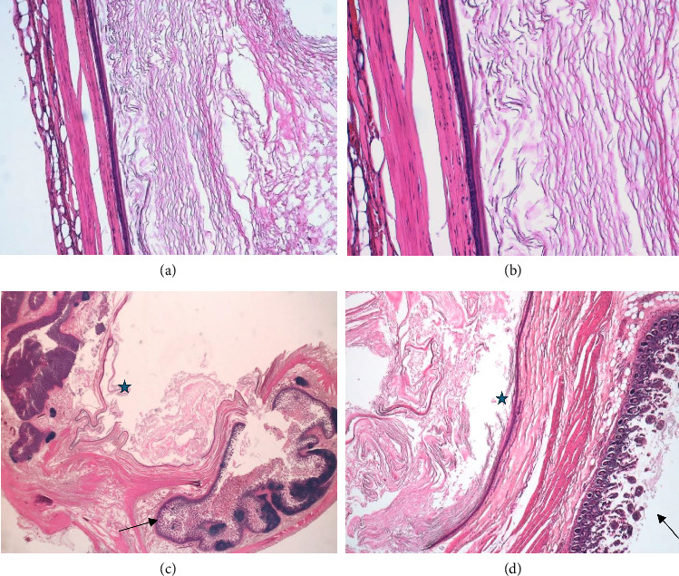 Figure 4
Peer Reviews
No public reviews on file for this paper yet. If you reviewed it on a platform where reviews are public (OpenReview, ICLR, NeurIPS, ICML), you can paste yours below so the community can read it here.
Videos
No videos yet. Explain this paper in a talk, walkthrough, or lecture? Add one.
Taxonomy
TopicsTeratomas and Epidermoid Cysts · Appendicitis Diagnosis and Management · Intraperitoneal and Appendiceal Malignancies
1. Introduction
Epidermoid cysts (ECs) are neoplasms that can develop in different parts of the body such as liver, spleen, kidney, and testis [1]. Those arising from the cecum are extremely rare; only 12 cases have been reported [2]. The origin can be acquired or congenital. Congenital EC is described in patients who do not have previous history of intra-abdominal surgery, abdominal trauma, or chronic inflammation; probably, it is the result of an aberrant ectodermal implantation during embryogenesis [3]. Conversely, the acquired form is associated with a history of abdominal surgery.
Basically, ECs are regarded as having benign characteristics, but a complete tumor resection is required due to the possibility of tumor recurrence or malignant transformation [4, 5].
Laparoscopic surgery has become the standard approach for colorectal surgery, offering shorter incisions, improved cosmesis, and better postoperative outcomes compared with an open approach [6].
We report a case of a 28-year-old woman, with no history of abdominal surgery or trauma, who was diagnosed with a cystic neoplasm of the cecum considered to meet the indications for laparoscopic surgery.
2. Case Presentation
A 28-year-old woman was admitted to the hospital with abdominal pain. She did not present prior history of chronic disease or previous surgeries. Vital signs were stable, no fever, vomiting or nausea were present. Physical examination revealed no tenderness in the abdomen, muscle guarding, or rebound tenderness. Laboratory data showed no inflammation or abnormal values.
Two abdominal magnetic resonance imaging (MRI) scans were performed, detecting a cystic mass of 104 × 83 × 68 mm in the anterior portion of the pelvic region. This neoplasm was close to the cecum without involving other abdominal organs such as uterus, bladder, sigma, and ovaries. The MRI scan showed a mass with hypoisointense signals on T1-weighted (T1W) images and hyperintense signal on T2-weighted (T2W) TSE images. Irregular heterogeneous areas of low signal intensity were noted in on both T1W and T2W images. Contrast-enhanced T1W images did not demonstrate enhancement. DWI showed high signal intensities (Figure 1). These MRI features can be observed both in ECs and in appendiceal mucocele [7]. However, the radiological assessment was initially misdiagnosed. The differential diagnosis included either a duplication cyst or an endometriotic cyst formation.
After 1 month, due to the worsening of the symptoms, an abdominal computed tomography (CT) scan was performed, confirming the presence of the cyst in close relation to cecum and ileum. This imaging investigation excluded appendicular mucocele as the appendix was normal in dimension and site (Figure 2).
Both CT and MRI are essential in ECs determinate the correct diagnosis and deciding on the surgical excision strategy [8]. CT scan showed an ovoid mass with fluid density (low attenuation), noninfiltrating, and well encapsulated with a sclerotic wall. The wall enhanced on contrast-enhanced images in the venous phase.
The case was discussed with a multidisciplinary equipped according to the indication for surgery. The procedure was executed with an explorative laparoscopy and the presence of a large neoplasm in the pelvic region was confirmed. This formation was attached to the cecum and the terminal ileum, without involving uterus and adnexa. Due to the nonspecific nature and the presence of numerous lymphadenopathies, a laparoscopic right hemicolectomy with an ileocolic stapled anastomosis was performed. The postoperative course was uneventful, and the patient was discharged on postoperative day 5.
2.1. Histopathological Diagnosis
The specimen was sent to the histopathology laboratory to be evaluated both macroscopically and microscopically.
The main macroscopic findings showed a 10 × 7 × 5 cm, whitish, round mass arising from the cecal wall. There was no apparent communication between the cyst and the mucosa of the cecum that appeared to be intact (Figure 3).
Microscopic examination revealed that the wall of the cyst was surrounded in its entirety by a cecal muscularis propria. The cyst wall was exclusively lined with a mature keratinized and stratified squamous epithelium with a granular layer, and focal area of cystic wall showed nonkeratinizing squamous epithelium without a granular layer. The lumen of the cyst was filled with mature, dense keratin (Figure 4).
The final histopathological diagnosis was the EC of the cecum.
3. Discussion
ECs of the cecum are extremely rare; however, similar cysts of internal organs have been reported involving the testis, epididymis, spleen, accessory spleen, kidney, and liver [1]. These cysts are generally accepted to be sequestration cysts with congenital or acquired origin.
Congenital ECs are related to inclusion of ectodermal elements at time of closure of neural groove or when epithelial surfaces fuse. Andiran et al. [9] suggested that the inclusion or closure line of the epidermal structure may occur when the cecum re-enters the abdominal cavity during intrauterine rotation in the final steps of gut development. This encasing of an epidermal structure may result in later development of a cecal epidermoid cyst (CEC). Acquired ECs are attributed to the iatrogenic implantation of epidermal fragments through surgical devices in previous abdominal operations, such as appendectomy or caesarean section [10, 11] or due to trauma.
Very few cases of EC of the cecum have been reported (Table 1).
Park et al. reported 9 cases including 3 cases (33.3%) of which are of acquired variety as have been associated with a history of abdominal surgery. Two had an appendectomy 12 years and 16 years before the diagnosis of a CEC [10, 11]. Furthermore, the case reported in 2012 was of a 31-year-old woman, with a previous caesarean surgery, who got admitted for an adnexal mass [14]. The other cases feature patients with no history of abdominal surgery, trauma, or chronic inflammation, and their CECs were considered as congenital lesions. Most of the reported cases in literature present patients with a history of chronic abdominal pain and other unspecific symptoms (nausea, asthenia, abdominal swelling, etc.) with the development of a palpable mass in the right lower abdominal quadrant. In some patients, ECs of the cecum are completely asymptomatic, and they are occasionally found during radiological exams performed for other reasons.
In our case, the patient had abdominal pain and MRI scans were performed detecting a cystic mass in the anterior portion of the pelvic region and after 1 month, and due to the worsening of the symptoms, an abdominal CT scan was performed, confirming the presence of the cyst. Physical examination and radiological findings are unspecific; in fact, none of the previous reports described a precise preoperative diagnosis. On CT, EC appears as a well-demarcated, low-density mass with enhancement of the capsule following contrast administration [18]. On MRI scan, the tumor appears hypointense on T1W imaging and hyperintense on T2W imaging.
The differential diagnosis is seldom taken into consideration in patients with cecal cystic lesions because ECs of the cecum are mainly located in the subserosal area [1, 3, 9, 12–14], as in our case. These cysts may be confused with other intra-abdominal cystic lesions, including appendiceal mucocele, duplication or mesenteric cyst, lymphatic cysts, gastrointestinal stromal tumors, and, in female patients, a right adnexal mass or cyst [2] or endometriotic cyst. Joo- Young Na et al. reported a case of congenital CEC in neonate initially misdiagnosed with retroperitoneal teratoma [17]. In the present case, the cystic mass showed on CT scan was suspected to be either an appendicular mucocele, a duplication cyst, or endometriotic cyst. Complete surgical excision of ECs is recommended to prevent recurrence and potential malignant transformation [19]. The residual tissue and cyst lining may cause recurrence; its rate has been reported as 2% [5]. In the present case, the laparoscopic right hemicolectomy with an ileocolic-stapled anastomosis was performed and the neoplasm was completely resected, as in cases of malignancy. Although there is no consensus in the literature regarding follow-up protocols after surgical resection of CECs, most authors suggest a long-term clinical and radiological surveillance, particularly in cases with uncertain margins or suspicion of malignant transformation.
In the literature, the laparoscopic approach was described as beneficial for both diagnosis and treatment of ECs. Only the most recent cases were treated by laparoscopic surgery, as in our case. This minimally invasive surgery procedure has recently become a popular approach to colon disease due to better short-term outcomes including reduced blood loss, improved intestinal function, and shorter duration of hospitalization [6]. Despite uncertainty about the origin of the mass, after the complete surgical excision has been performed, the histological examination of the specimen is the only investigation that provides a final diagnosis.
Macroscopically, these masses appear as solid neoplasms of variable size, up to 10 cm, as in our case. Microscopically, the cyst wall is lined by benign, keratinizing, stratified squamous epithelium with well-formed granular layer and abundant keratin but without skin specialized structures. The absence of skin specialized structures differentiates EC from dermoid cyst. There is no communication between the unilocular cyst and the normal colic mucosa.
4. Conclusions
ECs are rare and generally benign neoplasms. Several cases have been reported, showing a wide range of heterogeneity in both sex distribution and age.
Considering our case and literature review, the clinical presentation and the instrumental investigations can vary, making an immediate diagnosis difficult.
Therefore, the possibility of an EC should be taken into account in the differential diagnosis of submucosal or intramuscular masses in the cecal region.
The reference list from the paper itself. Each links out to its DOI / PubMed record.
- 1Uzunlar A. Bukte Y. Epidermoid Cyst of the Cecum in an Elderly Man Annals of Saudi Medicine 200626647747910.5144/0256-4947.2006.4772-s 2.0-3384668659717146207 PMC 6074325 · doi ↗ · pubmed ↗
- 2Park J. Y. Kim Y. W. Lee K. Y. Sung J. Y. Epidermoid Cyst of the Cecum Annals of Coloproctology 2015311373910.3393/ac.2015.31.1.372-s 2.0-8492586989025745626 PMC 4349916 · doi ↗ · pubmed ↗
- 3Sahoo M. R. Gowda M. S. Behera S. S. Unusual Site and Uncommon Presentation of Epidermoid Cyst: A Rare Case Report and Review of Literature BMJ Case Reports 20132013 p. bcr 201200790710.1136/bcr-2012-0079072-s 2.0-84872719654 PMC 360435423302551 · doi ↗ · pubmed ↗
- 4Kuroyanagi K. Kawabata T. Tooi M. Epidermoid Cyst of the Upper Lip: Report of a Case The Bulletin of Tokyo Dental College 197314295984516794 · pubmed ↗
- 5Ohsawa M. Kagawa T. Ochiai R. Rare Squamous Cell Carcinoma Arising From a Presacral Epidermoid Cyst: A Case Report International Journal of Surgery Case Reports 20206628328710.1016/j.ijscr.2019.12.02231884265 PMC 6939061 · doi ↗ · pubmed ↗
- 6Yamamoto S. Inomata M. Katayama H. JCOG 0404 Short-Term Surgical Outcomes From a Randomized Controlled Trial to Evaluate Laparoscopic and Open D 3 Dissection for Stage II/III Colon Cancer: Japan Clinical Oncology Group Study JCOG 0404 Annals of Surgery 20142601233010.1097/SLA.00000000000004992-s 2.0-8490225814624509190 · doi ↗ · pubmed ↗
- 7Hoang V. T. Trinh C. T. Nguyen C. H. Chansomphou V. Chansomphou V. Tran T. T. T. Overview of Epidermoid Cyst European Journal of Radiology Open 2019629130110.1016/j.ejro.2019.08.0032-s 2.0-8507165974531516916 PMC 6732711 · doi ↗ · pubmed ↗
- 8Radswiki T. Weerakkody Y. Sharma R. Appendiceal Mucocele Radiopaedia 202310.53347/r ID-12273 · doi ↗