An explainable unsupervised learning approach for anomaly detection on corneal in vivo confocal microscopy images
Ningning Tang, Qi Chen, Yunyu Meng, Daizai Lei, Li Jiang, Yikun Qin, Xiaojia Huang, Fen Tang, Shanshan Huang, Qianqian Lan, Qi Chen, Lijie Huang, Rushi Lan, Xipeng Pan, Huadeng Wang, Fan Xu, Wenjing He

TL;DR
This paper introduces a new AI method to detect corneal abnormalities in eye images without needing labeled examples of diseases.
Contribution
A Transformer-based unsupervised anomaly detection model for IVCM images with interpretable anomaly maps and improved generalizability.
Findings
The model achieved AUCs of 0.933 (internal) and 0.917 (external), outperforming existing methods.
Anomaly scores showed statistically significant differences between normal and pathological images (p < 0.001).
Explainable maps highlighted morphological deviations linked to potential corneal disease biomarkers.
Abstract
In vivo confocal microscopy (IVCM) is a crucial imaging modality for assessing corneal diseases, yet distinguishing pathological features from normal variations remains challenging due to the complex multi-layered corneal structure. Existing anomaly detection methods often struggle to generalize across diverse disease manifestations. To address these limitations, we propose a Transformer-based unsupervised anomaly detection method for IVCM images, capable of identifying corneal abnormalities without prior knowledge of specific disease features. Our method consists of three submodules: an EfficientNet network, a Multi-Scale Feature Fusion Network, and a Transformer Network. A total of 7,063 IVCM images (95 eyes) were included for analysis. The model was trained exclusively on normal IVCM images to capture and differentiate structural variations across four distinct corneal layers:…
Genes, proteins, chemicals, diseases, species, mutations and cell lines named across the full text — each resolved to its canonical identifier and authoritative record.
Click any figure to enlarge with its caption.
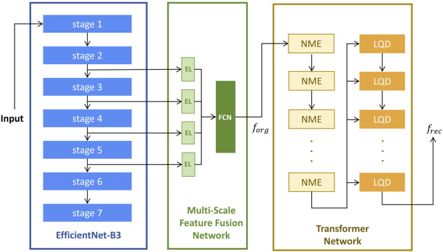 Figure 1
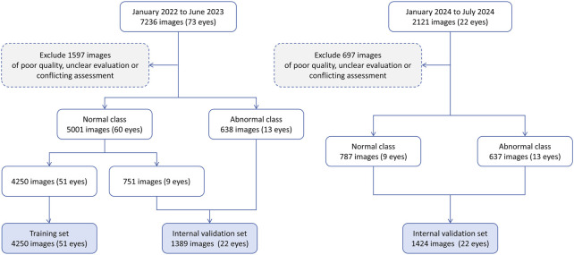 Figure 2
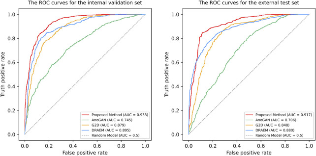 Figure 3
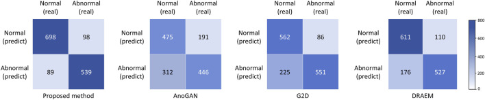 Figure 4
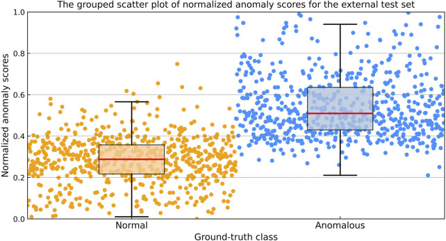 Figure 5
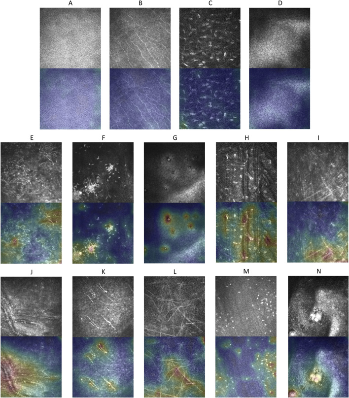 Figure 6
Peer Reviews
No public reviews on file for this paper yet. If you reviewed it on a platform where reviews are public (OpenReview, ICLR, NeurIPS, ICML), you can paste yours below so the community can read it here.
Videos
No videos yet. Explain this paper in a talk, walkthrough, or lecture? Add one.
Taxonomy
TopicsAnomaly Detection Techniques and Applications · Retinal Imaging and Analysis · Data-Driven Disease Surveillance