The parapharyngeal vein—an accessory communication between the middle cerebral veins and the internal jugular vein: a case report
Mugurel Constantin Rusu, Răzvan Costin Tudose, Alexandra Diana Vrapciu

TL;DR
This case report describes a rare anatomical variation where a parapharyngeal vein connects the middle cerebral veins to the internal jugular vein, offering a new understanding of venous drainage pathways.
Contribution
The paper identifies a previously unreported venous pathway involving the parapharyngeal vein as an accessory communication between cerebral and jugular veins.
Findings
A double superficial middle cerebral vein drained into the cavernous sinus and connected to the internal jugular vein via a parapharyngeal vein.
The parapharyngeal vein received tributaries like the facial and superior thyroid veins before draining into the internal jugular vein.
The absence of the right maxillary and retromandibular veins highlights the anatomical variability in this region.
Abstract
The superficial middle cerebral vein (SMCV) typically drains into the cavernous sinus, which, in turn, connects to the pterygoid venous plexus via a sphenoidal emissary vein. The latter may course through the foramen ovale. The pterygoid plexus drains in most cases into the retromandibular and facial veins. A peculiar SMCV drainage pathway to the internal jugular vein (IJV) via a parapharyngeal vein was found here. The anatomic variant reported here was identified by carefully reviewing the archived CT angiogram in a 68-year-old male case. A double SMCV was found on the right side. The resulting common SMCV trunk passed laterally to the foramen rotundum to empty into the cavernous sinus. A sphenoidal emissary vein joined it, which continued inferiorly through the foramen ovale to the pterygoid plexus. This plexus was connected to a reservoir on the inner side of the lateral pterygoid…
Genes, proteins, chemicals, diseases, species, mutations and cell lines named across the full text — each resolved to its canonical identifier and authoritative record.
Click any figure to enlarge with its caption.
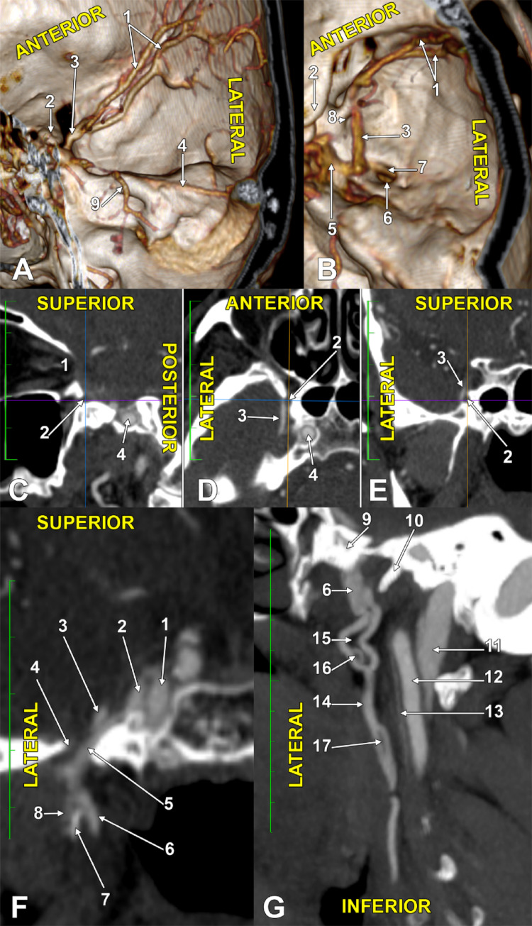 Figure 1
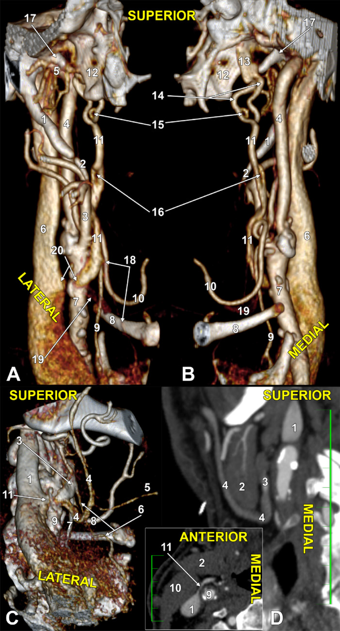 Figure 2
Peer Reviews
No public reviews on file for this paper yet. If you reviewed it on a platform where reviews are public (OpenReview, ICLR, NeurIPS, ICML), you can paste yours below so the community can read it here.
Videos
No videos yet. Explain this paper in a talk, walkthrough, or lecture? Add one.
Taxonomy
TopicsMeningioma and schwannoma management · Vascular Malformations Diagnosis and Treatment · Cerebral Venous Sinus Thrombosis