Self‐Assembly of Cholane–Phenanthrene–Cholane Trimers for Light‐Harvesting Supramolecular Systems
Edouard Ehret, Ioan Iacovache, Simon M. Langenegger, Benoît Zuber, Robert Häner

TL;DR
Researchers created light-harvesting nanostructures using cholesterol-based molecules, showing how their shape and efficiency depend on assembly conditions and additives.
Contribution
A new method for forming light-harvesting supramolecular systems using isomeric cholane–phenanthrene–cholane trimers.
Findings
Nanostructure morphology depends on phenanthrene isomer and cooling rate during assembly.
Doping with pyrene increases quantum yield fivefold, showing potential for artificial light-harvesting.
2D nanosheets and 3D worm-like or tubular structures are formed under different conditions.
Abstract
The self‐assembly of two isomeric amphiphilic cholane–phenanthrene–cholane trimers and the light‐harvesting properties of the formed supramolecular assemblies in aqueous medium are presented. Distinct differences in the supramolecular nanostructures are observed based on the type of phenanthrene isomer used, as well as on the cooling rate applied during the thermal self‐assembly process. A fast cooling rate results in the formation of 2D objects (nanosheets) with both trimers. In contrast, a slow cooling process results in the observation of 3D objects, with worm‐like nanostructures for the self‐assembled 3,6‐disubstituted phenanthrene trimer and nanotubes for the 2,7‐isomer. Upon doping with an acceptor chromophore, the supramolecular phenanthrene assemblies demonstrate efficient energy transfer. The presence of small quantities (6%) of a pyrene acceptor results in a fivefold increase…
Genes, proteins, chemicals, diseases, species, mutations and cell lines named across the full text — each resolved to its canonical identifier and authoritative record.
Click any figure to enlarge with its caption.
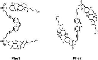 Figure 1
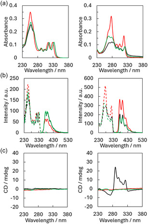 Figure 2
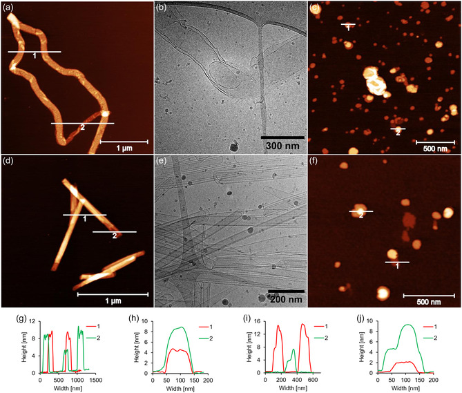 Figure 3
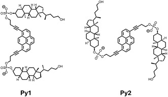 Figure 4
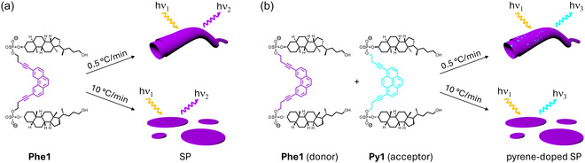 Figure 5
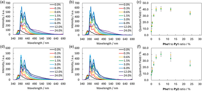 Figure 6
| Acceptor | Temperature gradient [°C min−1] | Quantum yield [%] | Increase in ΦF
|
|---|---|---|---|
|
| 0.5 | 41.9 ± 2.6 | 5× |
| 10 | 40.9 ± 3.7 | 5× | |
|
| 0.5 | 40.4 ± 3.2 | 5× |
| 10 | 40.8 ± 3.2 | 5× |
- —Schweizerischer Nationalfonds zur Förderung der Wissenschaftlichen Forschung 10.13039/501100001711
Peer Reviews
No public reviews on file for this paper yet. If you reviewed it on a platform where reviews are public (OpenReview, ICLR, NeurIPS, ICML), you can paste yours below so the community can read it here.
Videos
No videos yet. Explain this paper in a talk, walkthrough, or lecture? Add one.
Taxonomy
TopicsSupramolecular Self-Assembly in Materials · Synthesis and Properties of Aromatic Compounds · Metal-Organic Frameworks: Synthesis and Applications
Introduction
1
The development of self‐assembling supramolecular systems represents a cornerstone of modern materials science, offering unprecedented opportunities to design functional nanomaterials.^[^ 1, 2, 3, 4, 5, 6, 7, 8 ^]^ These materials are composed of repeating molecular units held together by noncovalent interactions, enabling precise structural and functional control while retaining dynamic reversibility.^[^ 9, 10, 11, 12, 13, 14, 15, 16, 17 ^]^ Among the various approaches, amphiphilic, π‐conjugated building blocks have proven particularly effective for the assembly of nanostructures with tailored properties for applications ranging from drug delivery to optoelectronics and light‐harvesting complexes (LHCs).^[^ 18, 19, 20, 21, 22, 23, 24, 25, 26, 27, 28 ^]^ In LHCs, efficient energy transfer depends on the ordered positioning of chromophores, which self‐assembly facilitates by allowing precise spatial arrangements and fine‐tuning of their organization.^[^ 29, 30, 31 ^]^ Moreover, the ability to modify the individual building block with different chromophores makes supramolecular polymers (SPs) ideal candidates for energy transfer systems, light‐harvesting devices, or advanced sensing technologies. Previously, we reported on the self‐assembly of amphiphilic, π‐conjugated, phosphodiester‐linked trimers in aqueous media.^[^ 32, 33, 34 ^]^ Upon a controlled cooling step, these compounds were shown to form supramolecular assemblies with various morphologies ranging from nanosheets to nanotubes and nanoribbons. Additionally, such assemblies were shown to possess impressive light‐harvesting properties.^[^ 35, 36 ^]^
In this work, we report the synthesis of two phosphodiester‐linked cholane–phenanthrene–cholane trimers bearing two different substitution patterns (Phe1 and Phe2, Figure 1) and their self‐assembly, as well as light‐harvesting properties in the presence of a suitable acceptor chromophore. While the phenanthrene units act as chromophores, the hydrophobic cholanes promote self‐assembly in an aqueous environment. Doping of the self‐assembled nanostructures with a pyrene chromophore resulted in an efficient energy transfer process.
The two cholane‐phenanthrene‐cholane trimers Phe1 and Phe2 used in this study.
Results and Discussion
2
Supramolecular Assembly of Cholane–Phenanthrene–Cholane Trimers into Different Types of Morphologies
2.1
The synthesis of Phe1 and Phe2 was performed by adapting published procedures.^[^ 33, 35 ^]^ Lithocholic acid was reduced to its diol form and selectively protected. Subsequently, it was coupled to its phenanthrene diphosphoramidite. The trimers were deprotected, purified by RP‐HPLC, and characterised by HRMS (see Supporting Information).
Temperature‐dependent UV‐vis absorption spectra were recorded for both trimers to investigate their self‐assembly process (Figure 2a). To ensure complete disassembly, all samples were initially heated to 70 °C and then cooled to 20 °C with two different rates: 0.5 °C min^−1^ (slow) and 10 °C min^−1^ (fast). During the cooling process (70–20 °C), characteristic changes were observed in the absorbance of the two trimers, indicating the self‐assembly of the SPs. For Phe1, the spectrum showed a bathochromic shift for the bands between 314 and 328 nm accompanied by a hypochromic effect at 257 nm at 20 °C after applying the slow cooling gradient (black curve). Similarly, for Phe2, a pronounced hypochromicity was evident across the spectrum, along with a 3 nm red shift for bands between 294 and 318 nm. The self‐assembly process of both trimers followed a nucleation‐elongation process which starts around 51 °C (Figure S18, Supporting Information).^[^ 14, 37 ^]^ The change to a faster cooling rate did not have a significant influence on the absorption by the self‐assembly formed by Phe1 at 20 °C (green curve). It did, however, induce changes in the absorption by the self‐assembled Phe2, as observed by the smaller hypochromic effect present in the region between 260 nm and 290 nm. Further characterization of the assemblies involved measuring temperature‐dependent excitation and emission fluorescence spectra (Figure 2b). Excitation of Phe1 at 333 nm revealed an increase in the phenanthrene monomer emission upon self‐assembly regardless of the temperature gradient applied. In contrast, Phe2 exhibited a strong decrease in phenanthrene monomer fluorescence upon cooling, with a more pronounced reduction under the fast cooling gradient. Furthermore, circular dichroism (CD) spectra were recorded before and after the self‐assembly process (Figure 2c). In its assembled state, Phe1 did not show any CD signal after either cooling condition. However, Phe2 displayed dichroic signals after self‐assembly using the slow cooling gradient, as indicated by the black curve. This effect was absent when the fast cooling gradient was applied. The changes observed in the UV‐vis and fluorescence spectra suggest that an aggregation process is occurring for both trimers and that this process differs for Phe2 depending on the cooling rate applied.
Spectroscopic characterization of Phe1 (left) and Phe2 (right). The spectra were measured at 70 °C (red) and 20 °C, after slow cooling (black) and after fast cooling (green). a) Temperature‐dependent UV‐vis absorption spectra; b) fluorescence excitation spectra (dotted line) and fluorescence emission spectra (solid line) left: λex.: 333 nm and λem.: 375 nm, right: λex.: 320 nm and λem.: 394 nm; c) CD spectra. Conditions: 3 μM oligomer, 10 mM sodium phosphate buffer pH 7.2, 10 mM NaCl, and EtOH (15% for Phe1 and 20% for Phe2).
The morphologies of the self‐assembled aggregates were studied by atomic force microscopy (AFM) and cryo‐electron microscopy (cryo‐EM, Figures 3 and S21–S26, Supporting Information). After assembly, the samples were deposited onto either (3‐aminopropyl)triethoxysilane (APTES)‐modified mica sheets for AFM or on copper lacey carbon grids for cryo‐EM. Despite exhibiting identical spectroscopic properties under two different cooling gradients, Phe1 self‐assembled into two distinct supramolecular nanostructures depending on the cooling rate. AFM and cryo‐EM measurements revealed worm‐like nanostructures formed at a slow cooling gradient (Figure 3a,b), while circular nanosheets were observed at a fast cooling rate (Figure 3c). Cryo‐EM images show that the worm‐like structures are between 100 and 310 nm wide, up to 30 μm long, and possess a double membrane. AFM cross sections indicate heights of 4 to 8 nm suggesting a membrane thickness of ≈2 nm. The observed nanosheets have a height of 4 or 8 nm and possess a diameter varying from 50 to 150 nm. In contrast, Phe2, the 2,7‐disubstituted trimer, self‐assembled into nanotubes at a slow cooling rate (Figure 3d,e) and into nanosheets at a fast rate (Figure 3f). Cryo‐EM measurements show the presence of single‐ and multi‐walled nanotubes, with widths ranging from 30 to 80 nm and up to 2 μm long. AFM imaging measurements correlate well with these findings, showing nanotube heights ranging from 4 to 16 nm, with 4 nm increments. This indicates a membrane thickness of about 2 nm. The nanosheets formed by this phenanthrene isomer were thinner than those formed by Phe1, with a height of 2 nm and a diameter ranging from 50 to 200 nm. Thus, in addition to the molecular structure of the phenanthrene units, the assembly process is also significantly affected by the cooling rate.^[^ 38 ^]^
Microscopy measurements of oligomers Phe1 (top) and Phe2 (bottom). AFM images of the oligomers after a cooling rate of a,d) 0.5 °C min−1; c,f) 10 °C min−1; b,e) cryo‐EM images of the oligomers after cooling (0.5 °C min−1); g–j) cross sections of the AFM images a, c, d, and f, respectively. Conditions: 3 μM oligomer, 10 mM sodium phosphate buffer pH 7.2, 10 mM NaCl, and EtOH (15% for Phe1; 20% for Phe2).
Light‐Harvesting Properties of Phe1
2.2
Next, we studied the light‐harvesting properties of the Phe1‐based SPs. For this purpose, the assembly of Phe1 was performed in the presence of small quantities of a cholane–pyrene–cholane trimer (Py1 or Py2, Figure 4). The pyrene‐containing trimers are incorporated into the growing SPs during the assembly process and can act as acceptor chromophores after excitation of phenanthrene.^[^ 39 ^]^ Thus, pyrene‐doped SPs were assembled following different temperature gradients (0.5 and 10 °C min^−1^) as described above. An illustration of the formation of the SP via self‐assembly of Phe1 in the absence of Py1 is shown in Figure 5a, as well as in the presence of Py1 in Figure 5b. Similar results were obtained in light‐harvesting experiments using Phe2. For simplicity, only the data acquired with Phe1 are described.
Chemical structures of both acceptor oligomers used as doping agents.
Illustration of an idealized assembly of an SP of Phe1 (purple) in the absence a) or presence b) of an acceptor oligomer (blue), which is randomly integrated into the forming SP. For clarity, only one acceptor oligomer (Py1) is indicated here, but equal results are obtained with Py2.
The light‐harvesting properties of the supramolecular nanostructures formed by Phe1 were determined by recording fluorescence spectra (Figure 6) in the presence of up to 24% of Py1 (top) or Py2 (bottom). Selective excitation of Phe1 at 333 nm leads to major changes in the fluorescence emission spectrum of the system at 20 °C after slow and fast cooling rates (Figure 6a,b, respectively). The gradual increase in the amount of acceptor chromophore leads to a decrease in phenanthrene emission (max. at 376 nm, black line) while the pyrene monomer emission (four maxima at 393, 399, 419 and 440 nm) reaches maximum intensity after the addition of 6% of Py1 before decreasing again. In addition, the addition of larger quantities of Py1 (24%) leads to the appearance of a broad band (from 460 to 580 nm). This band is attributed to pyrene excimer formation, since rising concentrations of pyrene molecules increase the probability of molecular interaction.^[^ 40 ^]^ These changes are in correlation with the fluorescence quantum yield (Φ_F_, Figure 6c) of the system, which increases from 8.3 ± 0.6% to a maximum of 41.9% ± 2.6% at 6% of Py1, followed by a slight decrease. The cooling rate applied to the system did not have a significant effect on the fluorescence properties as can be seen in Figure 6c. The quantum yields are equal (Table 1) for SPs obtained by applying either of the temperature gradients. Similar results were obtained after the addition of Py2 with the appearance of pyrene monomer emission (three maxima at 396, 418 and 440 nm) after slow and fast cooling rates (Figure 6d,e, respectively). Also in this case, the addition of increasing amounts of Py2 results in a decrease in the phenanthrene emission and an increase in the pyrene monomer emission. A maximum in fluorescence emission is reached after the addition of 6% of Py2, while a distinct pyrene excimer emission band is present after the addition of 12–24% of the acceptor. The fluorescence quantum yield (Figure 6f) reaches a maximum of 40.4 ± 3.2% after the addition of 6% of Py2 (Table 1), which corresponds to a fivefold increase compared to the system in the absence of an acceptor. AFM measurements performed on doped systems in the presence of 6% of the acceptor trimer (see Figures S37 and S38, Supporting Information) revealed that the presence of the acceptor did not influence the morphology of the different SPs.
Fluorescence emission spectra of Phe1 in the presence of increasing amounts of Py1 (top) and Py2 (bottom) (0 to 24%) with a cooling rate of a,d) 0.5 °C min−1 and b,e) 10 °C min−1. c,f) Quantum yield of both systems, in green at 0.5 °C min−1 and in yellow at 10 °C min−1. Conditions: 3 μM Phe1, 10 mM sodium phosphate buffer pH 7.2, 10 mM NaCl, and 15% EtOH, λex: 333 nm.
The observations described above indicate that an energy transfer process takes place between phenanthrene and pyrene molecules. The decrease in fluorescence of the donor (phenanthrene) in the presence of the acceptor (pyrene) can be explained by a FRET mechanism.^[^ 41, 42 ^]^ However, the increase in the quantum yields indicates that additional energy transfer mechanisms are also involved (e.g., coherent energy transfer).^[^ 43, 44, 45 ^]^
Interestingly, the difference in the supramolecular morphology did not have any influence on the light‐harvesting capabilities of the Phe1 donor trimer as highlighted by the nearly identical quantum yields observed after both cooling rates were applied (see Table 1).
Conclusions
3
The synthesis and self‐assembly processes of two isomeric phosphodiester‐linked cholane–phenanthrene–cholane trimers have been described. AFM and cryo‐EM measurements revealed different types of morphologies depending on the phenanthrene isomer used and the cooling gradient applied during a thermally controlled self‐assembly process. For Phe1, a cooling rate of 0.5 °C min^−1^ resulted in the formation of worm‐like nanostructures. Yet, applying a cooling rate of 10 °C min^−1^ resulted in the formation of supramolecular nanosheets. Furthermore, the second isomer investigated, Phe2, self‐assembled into single‐ and multi‐walled nanotubes when a cooling rate of 0.5 °C min^−1^ was applied. Finally, using a faster cooling gradient (10 °C min^−1^), the self‐assembly of Phe2 resulted in the formation of nanosheets.
In addition, the supramolecular assemblies of the 3,6‐dialkynyl phenanthrene trimer (Phe1) revealed remarkable light‐harvesting properties. Doping of the supramolecular assembly with a pyrene acceptor chromophore leads to an efficient energy transfer between phenanthrenes and the pyrenes, regardless of the morphology (worm‐like nanostructure or nanosheet). Thus, the addition of 6% of Py1 or Py2 resulted in a fluorescence quantum yield of 40%, which corresponds to a fivefold increase compared to the native (undoped) Phe1. These data demonstrate that the supramolecular assembly of short, amphiphilic oligomers has a positive effect on the energy transfer processes between the phenanthrene and pyrene chromophores. In view of the simplicity of the assembly process and their compatibility with aqueous conditions, cholane–phenanthrene–cholane phosphodiester‐linked compounds present promising building blocks for artificial LHCs and other functional supramolecular systems.
Conflict of Interest
The authors declare no conflict of interest.
Supporting information
Supplementary Material
The reference list from the paper itself. Each links out to its DOI / PubMed record.
- 1G. R. Whittell , M. D. Hager , U. S. Schubert , I. Manners , Nat. Mater. 2011, 10, 176.21336298 10.1038/nmat 2966 · doi ↗ · pubmed ↗
- 2J.‐F. Lutz , J.‐M. Lehn , E. W. Meijer , K. Matyjaszewski , Nat. Rev. Mater. 2016, 1, 16024.
- 3P. M. G. Löffler , O. Ries , A. Rabe , A. H. Okholm , R. P. Thomsen , J. Kjems , S. Vogel , Angew. Chem., Int. Ed. 2017, 56, 13228.10.1002/anie.20170324328598002 · doi ↗ · pubmed ↗
- 4J. L. Greenfield , J. R. Nitschke , Acc. Chem. Res. 2022, 55, 391.35015510 10.1021/acs.accounts.1c 00657 · doi ↗ · pubmed ↗
- 5D. Gupta , B. S. Varghese , M. Suresh , C. Panwar , T. K. Gupta , J. Nanopart. Res. 2022, 24, 196.
- 6L. Ge , H. Xu , X. Jiang , J. Yu , CCS Chem. 2023, 6, 69.
- 7M. Harun‐Ur‐Rashid , I. Jahan , Md. J. Islam , A. Kumer , M. N. Huda , A. B. Imran , S. Gouadria , S. A. Alsalhi , J. Mol. Struct. 2024, 1304, 137665.
- 8X. Dang , Q. Zhang , F. Cui , T. Xiao , Chem Photo Chem 2025, 1324, e 202400392.